Review
. 2009 Jan;457(3):609-22.
doi: 10.1007/s00424-008-0495-4. Epub 2008 Jun 6.

The gastric HK-ATPase: structure, function, and inhibition

Affiliations
Review

The gastric HK-ATPase: structure, function, and inhibition

Jai Moo Shin et al. Pflugers Arch. 2009 Jan.

Erratum in

  • Pflugers Arch. 2011 Mar;461(3):399

Abstract

The gastric H,K-ATPase, a member of the P(2)-type ATPase family, is the integral membrane protein responsible for gastric acid secretion. It is an alpha,beta-heterodimeric enzyme that exchanges cytoplasmic hydronium with extracellular potassium. The catalytic alpha subunit has ten transmembrane segments with a cluster of intramembranal carboxylic amino acids located in the middle of the transmembrane segments TM4, TM5,TM6, and TM8. Comparison to the known structure of the SERCA pump, mutagenesis, and molecular modeling has identified these as constituents of the ion binding domain. The beta subunit has one transmembrane segment with N terminus in cytoplasmic region. The extracellular domain of the beta subunit contains six or seven N-linked glycosylation sites. N-glycosylation is important for the enzyme assembly, maturation, and sorting. The enzyme pumps acid by a series of conformational changes from an E(1) (ion site in) to an E(2) (ion site out) configuration following binding of MgATP and phosphorylation. Several experimental observations support the hypothesis that expulsion of the proton at 160 mM (pH 0.8) results from movement of lysine 791 into the ion binding site in the E(2)P configuration. Potassium access from the lumen depends on activation of a K and Cl conductance via a KCNQ1/KCNE2 complex and Clic6. K movement through the luminal channel in E(2)P is proposed to displace the lysine along with dephosphorylation to return the enzyme to the E(1) configuration. This enzyme is inhibited by the unique proton pump inhibitor class of drug, allowing therapy of acid-related diseases.


Figures

Fig. 1
Fig. 1
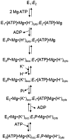
The catalytic cycle of the gastric H,K-ATPase. A hydronium ion binds to the cytoplasmic surface of the enzyme, and MgATP phosphorylates the protein at Asp386 to form the first ion transport intermediate in the E1 form. E1 form then converts by a conformational change to the second ion transport form, E2, with the ion site now exposed to the exterior and hydronium is released at pH ~1.0. To this form, K+ binds from the outside surface to the same region from which the hydronium was released, and the enzyme dephosphorylates, and then K+ is trapped within the membrane domain in what was called the occluded form (and a similar form is postulated for the hydronium in the outward step of the cycle). The K+ is then de-occluded, allowing reformation of the E1 form of the enzyme with the ion site now again facing the cytoplasm, and K+ is displaced when ATP is bound. The hydronium ion (H3O+) is used instead of the naked H+, as this enables the necessary hydrogen bonding and is more akin to Na+ that is transported by the H,K-ATPase at high pH. The above reflects a stoichiometry of 1H/1K/1ATP
Fig. 2
Fig. 2
Ribbon diagram depicting the ion entry and exit pathways of the gastric H,K-ATPase: The upper left image illustrates the conformation of the pump in the E1P form, and the intramembranal carboxylic acids are shown in stick form and numbered in blue, with the arrow highlighting the position of lysine791. Three hydronium ions are shown in binding sites designated H1, H2, H3. The upper right image illustrates the E2P conformation where lysine 791 has displaced the hydronium in H2 to the luminal face, and an arrow now emphasizes the new orientation of lysine 791. Large changes in orientation of the first four transmembrane segments, M1 (dark blue ribbon), M2 (light blue ribbon), M3 and M4 (green ribbon), generate the ion path for hydronium exit and K+ entry in E2P as well as a luminal vestibule leading to the pathway (bottom of the lower figure). The bottom image illustrates the entry path for K+ (illustrated as a series of violet spheres) between M4, M5, M6, and M8 in the E2P conformation as determined by molecular dynamics simulation (M3 in the foreground is omitted for clarity). The arrival of K+ at the top of this path is predicted to destabilize the interaction of lysine 791 with E820 and E795 and initiate the conformational changes leading to release of phosphate at the active site and conversion back to E1. Not shown is the postulated movement of K+ into the position of the third hydronium associated with glutamyl 343 before returning to the cytoplasmic face of the enzyme at the top of the respective illustrations
Fig. 3
Fig. 3
General changes in the orientation and shape of the gastric H,K-ATPase in the transition from the E1 to the E2 conformation. On the left is shown the N, P, and A domains before phosphate transfer from MgATP where the three cytoplasmic domains change conformation, with the A, N, and P domains now closer in the E1P form and a further change also in the membrane domain with the formation of the E2P form that allows expulsion of a proton into the exoplasmic region to be followed by K uptake
Fig. 4
Fig. 4
A comparison of the ion site structure in E2P of the SERCA Ca-ATPase and the gastric H,K ATPase showing the need for expansion of the ion site due to the insertion of the NH3+ group of lysine 791 to energize expulsion of the proton from the E2P form of the gastric ATPase
Fig. 5
Fig. 5
The reactions catalyzed by the oligomeric form of the gastric H,K-ATPase where one heterodimer undergoes the cycle from E1 to E2 as illustrated in Fig. 1, while the other (italics) adopts a reciprocal conformation, i.e., E2 corresponding to E1 and E1 corresponding to E2 in the two out of phase oligomers. One of the heterodimers is italicized
Fig. 6
Fig. 6
Different proton pump inhibitors
Fig. 7
Fig. 7
The rate constants of activation of the different PPIs as a function of decreasing medium pH. It can be seen that they are slowly activated at a pH >3.0, but then activation increases rapidly at lower pH values with lansoprazole slightly faster than omeprazole and pantoprazole slightly faster than tenatoprazole but the latter two clearly slower than the former. The rate of activation for lansoprazole decreases at pH values less than 0
Fig. 8
Fig. 8
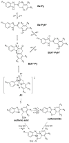
The mechanism of activation of the PPIs shown in general structural form. The top of the figure shows the protonation of the pyridine ring, and the second row of structures shows protonation also of the benzimidazole ring. The bis-protonated forms are in equilibrium with the protonated benzimidazole and unprotonated pyridine. In brackets is shown the mechanism of activation whereby the 2C of the protonated benzimidazole reacts with the unprotonated fraction of the pyridine moiety that results in rearrangement to a permanent cationic tetracyclic sulfenic acid which, in aqueous solute, dehydrates to form a cationic sulfenamide. Either of these thiophilic species can react with the enzyme to form disulfides with one or more enzyme cysteines accessible from the luminal surface of the enzyme
Fig. 9
Fig. 9
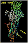
The binding sites of pantoprazole to the H,K-ATPase showing one site at cysteine 813 in the vestibule of the pump, and cysteine 822 2.5 turns into the membrane domain in TM6
Fig. 10
Fig. 10
The rate of loss of bound PPI (PNZ pantoprazole, OPZ omeprazole) as a function of time of incubation with 10 mM glutathione. The removal of labeled drug is biphasic, the fast phase accounts for about 84% of omeprazole binding and 60% of pantoprazole labeling, whereas the slow phase represents about 16% of omeprazole labeling and 40% of pantoprazole labeling
Fig. 11
Fig. 11
The labeling of the ATPase in vivo as a function of time after IV administration of pantoprazole (PNZ) or omeprazole (OPZ). It can be seen that the rate of loss of omeprazole is about twice that of pantoprazole and that the stoichiometry of labeling for almost full inhibition of enzyme is about 2.6 nmol drug bound per milligram H, K-ATPase. The error bars represent ±SEM, n=4–5 animals in each group
Fig. 12
Fig. 12
Imidazopyridine core structures of some acid pump antagonists
Fig. 13
Fig. 13
Different views of the K+-competitive inhibitor BYK36399 binding site from within the plane of the membrane in the first two images. The residues affecting inhibitor binding in TM4 are shown in green and in the TM5/6 loop are shown in yellow and in TM6 in gold. On the far right is shown the docking of the methylated form, N-methyl BYK36399, where the affinity is much reduced due to steric hindrance (arrows) with Ala 335 and Cys 813. The proximity of Cys 813, a common binding site for the PPIs, to the calculated structure for binding of the APA should be noted. The largest changes in Ki were generated by the mutants Ala335Cys, Phe331Ile, and Leu809Phe. This docking model is also consistent with the binding of an azido derivative of SCH28080 to the region of TM1 and TM2 [31, 34]

References

    1. Abe K, Kaya S, Taniguchi K, Hayashi Y, Imagawa T, Kikumoto M, Oiwa K, Sakaguchi K. Evidence for a relationship between activity and the tetraprotomeric assembly of solubilized pig gastric H/K-ATPase. J Biochem (Tokyo) 2005;138:293–301. - PubMed
    1. Andersson K, Carlsson E. Potassium-competitive acid blockade: a new therapeutic strategy in acid-related diseases. Pharmacol Ther. 2005;108:294–307. - PubMed
    1. Asano S, Yoshida A, Yashiro H, Kobayashi Y, Morisato A, Ogawa H, Takeguchi N, Morii M. The cavity structure for docking the K(+)-competitive inhibitors in the gastric proton pump. J Biol Chem. 2004;279:13968–13975. - PubMed
    1. Bamberg K, Mercier F, Reuben MA, Kobayashi Y, Munson KB, Sachs G. cDNA cloning and membrane topology of the rabbit gastric H+/K(+)-ATPase alpha-subunit. Biochim Biophys Acta. 1992;1131:69–77. - PubMed
    1. Besancon M, Shin JM, Mercier F, Munson K, Miller M, Hersey S, Sachs G. Membrane topology and omeprazole labeling of the gastric H+,K(+)-adenosinetriphosphatase. Biochemistry. 1993;32:2345–2355. - PubMed

Publication types

Substances

LinkOut - more resources