. 2008 Sep;213(3):336-41.
doi: 10.1111/j.1469-7580.2008.00919.x. Epub 2008 Jun 5.

Compliance in the neck structures of the guinea pig spermatozoon, as indicated by rapid freezing and electron microscopy

Affiliations

Compliance in the neck structures of the guinea pig spermatozoon, as indicated by rapid freezing and electron microscopy

D M Woolley et al. J Anat. 2008 Sep.

Abstract

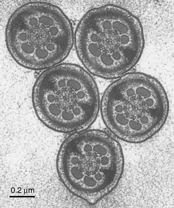
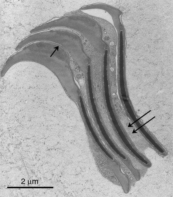
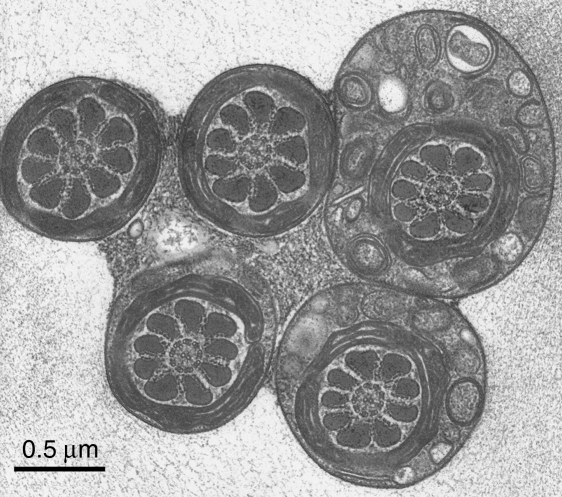
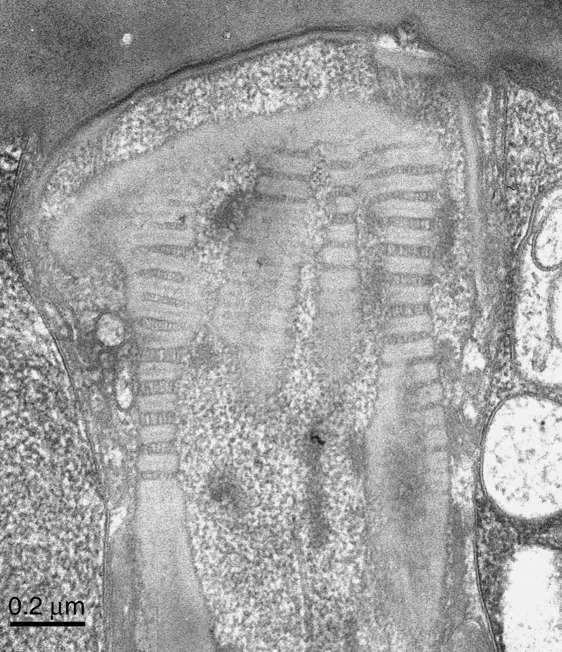
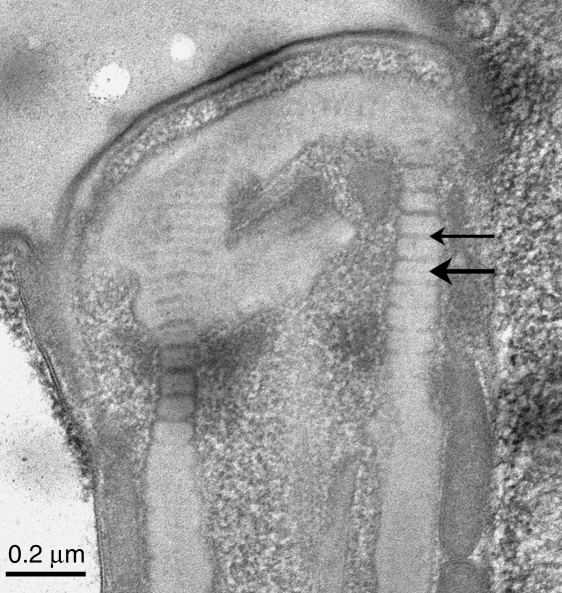
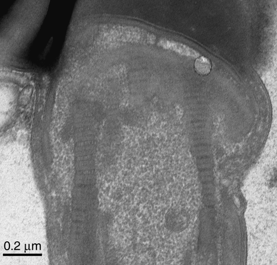
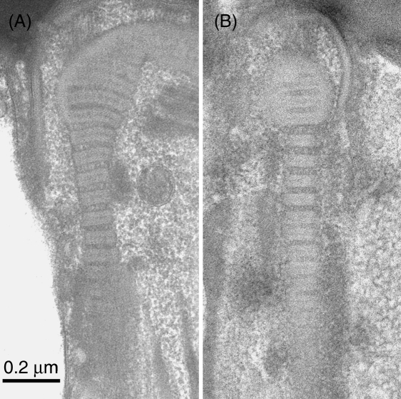
Electron microscopy has been used to investigate whether the transversely striated columns of the connecting piece in the neck region of guinea pig spermatozoa, undergo lengthening and shortening as a result of the forces generated during motility. Motile spermatozoa were subjected to near-instantaneous rapid freezing, followed by freeze-substitution fixation and epoxy embedment. Thin sections passing longitudinally through the striated columns revealed that the periodicity was indeed variable. The repeat period, taken to have an unstressed width of 60 nm, could be found extended to 75 nm in some specimens, and reduced to 54 nm in others. The estimates of the coefficients of variation were 6.6% for the width of the 'dense' band and 33.5% for the 'pale' band. The 'pale' band in the extended state showed longitudinal striae. Such variations in length, which - it is suggested - are physiological, and passively induced, would have functional implications for the flagellum - for both bend initiation and bend growth. Also, hypothetically, any mechanism that could increase the degree of compliance in these columns, such as perhaps phosphorylation of the constituent proteins, could permit the flagellum to develop the exaggerated bend angles and asymmetries of the 'hyperactivated' state.


Figures

Fig. 2
Fig. 2
A group of sperm flagella sectioned at the level of the principal piece, from a thin section cut perpendicularly to the plane of rapid freezing. As expected from the nature of the head rouleau, groups of tails such as this are always similarly orientated. The axoneme itself has the standard arrangement, without any obvious dynamic variations in the interdoublet spacings. Scale bar=0.2µm.
Fig. 1
Fig. 1
A demonstration that the normal ‘rouleaux’ formation of the spermatozoa is preserved throughout the slam-freezing and freeze-substitution procedure. This rouleau comprises four sperm sectioned at the level of the acrosome. The heads are linked together by the well known membrane appositions (arrowed) – see Friend & Fawcett (1974). However, granular material, containing vesicles, is also present between the sperm heads (double arrows); this material is scarcely preserved at all in conventionally fixed cells (loc. cit.). Scale bar=2µm.
Fig. 3
Fig. 3
A group of sperm flagella sectioned at the level of the midpiece, from a thin section cut perpendicularly to the freezing plane. At this level the flagella are stuck together by the granular material, which has been shown to lie also between the sperm heads (Fig. 1). This mechanical coupling must have a role in the flagellar synchronization – which is generally explained in the literature on cilia and flagella as being due to viscous coupling. Again, the structure of the midpiece and of the cytoplasmic droplet (top right) is well preserved and conventional. Scale bar=0.5µm.
Fig. 5
Fig. 5
This micrograph was taken a few minutes before that in Fig. 4, that is, at the same instrumental settings. Here the periodicities within the columns are highly variable. It is evident that most of the variation is in the ‘pale’ bands, which in places are increased in width to equal the width of the ‘dense’ bands’. Where this is so, for example at the lower left, the ‘pale’ bands show longitudinal striae. The variations in period are analysed further in the text. Scale bar=0.2µm.
Fig. 4
Fig. 4
A longitudinal section through the connecting piece. This and all subsequent specimens are from sections cut parallel to the freezing plane. This example resembles the pattern seen after conventional fixation except that the contrast is reversed – for unknown reasons. Thus, the normally dense and wide bands of the striated columns appear pale (large arrow) and the normally pale and thin bands appear dense (small arrow). (For continuity, the bands are nevertheless referred to as ‘dense’ and ‘pale’, respectively.) The periodicities in this specimen were rather uniform. The mean repeat periods, as measured, were 60.6nm (left side) and 59.0nm (right side). Scale bar=0.2µm.
Fig. 7
Fig. 7
This micrograph demonstrates within-sperm variation, with the periodicity of the right side column (mean width 64.4nm) less than that on the left side (74.1nm). Scale bar=0.2µm.
Fig. 6
Fig. 6
(A, B) These two examples show that the periodicity in the columns may be extended in the middle regions but is at a standard width in the distal region, where the column abuts the origin of the outer dense fibre. This suggests that there is no appreciable compliance in the substance of the latter. Scale bar=0.2µm.

References

    1. Bremser J. Gametes and Spores. Baltimore: Johns Hopkins University Press; 1819. (drawings re-published by J Farley (1982)) p. 45.
    1. Chan PJ, Corselli JU, Patton WC, Jacobson JD, King A. Enhanced fertility after heat-induced hyperactivation. Fertil Steril. 1998;69:118–121. - PubMed
    1. Cody BA. Observations and experiments upon spermatozoa of the guinea pig. J Urol. 1925;13:175–191.
    1. Fawcett DW. The anatomy of the mammalian spermatozoon with particular reference to the guinea pig. Z Zellforsch Mikrosk Anat. 1965;67:279–296. - PubMed
    1. Fawcett DW, Phillips DM. The fine structure and development of the neck region of the mammalian spermatozoon. Anat Rec. 1969;165:153–184. - PubMed

LinkOut - more resources