

Utility of biomarkers in prediction of response to ablative therapy in Barrett's esophagus
- PMID: 18538141
- PMCID: PMC3896328
- DOI: 10.1053/j.gastro.2008.04.036
Utility of biomarkers in prediction of response to ablative therapy in Barrett's esophagus
Abstract
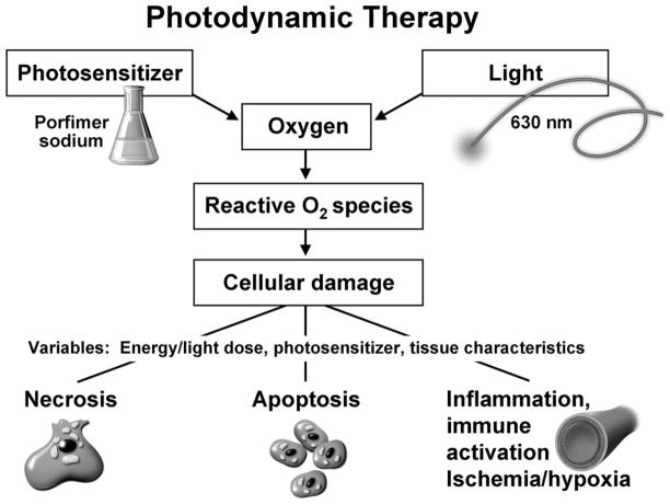
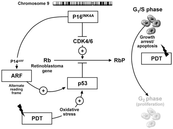
Background & aims: Photodynamic therapy (PDT) has been shown to be effective in the treatment of high-grade dysplasia (HGD)/mucosal carcinoma in Barrett's esophagus (BE). Substantial proportions of patients do not respond to PDT or progress to carcinoma despite PDT. The role of biomarkers in predicting response to PDT is unknown. We aimed to determine if biomarkers known to be associated with neoplasia in BE can predict loss of dysplasia in patients treated with ablative therapy for HGD/intramucosal cancer.
Methods: Patients with BE and HGD/intramucosal cancer were studied prospectively from 2002 to 2006. Biomarkers were assessed using fluorescence in situ hybridization performed on cytology specimens, for region-specific and centromeric probes. Patients were treated with PDT using cylindric diffusing fibers (wavelength, 630 nm; energy, 200 J/cm fiber). Univariate and multiple variable logistic regression was performed to determine predictors of response to PDT.
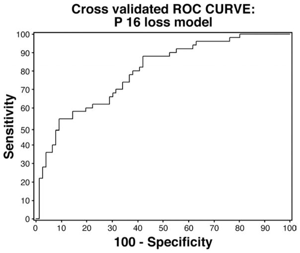
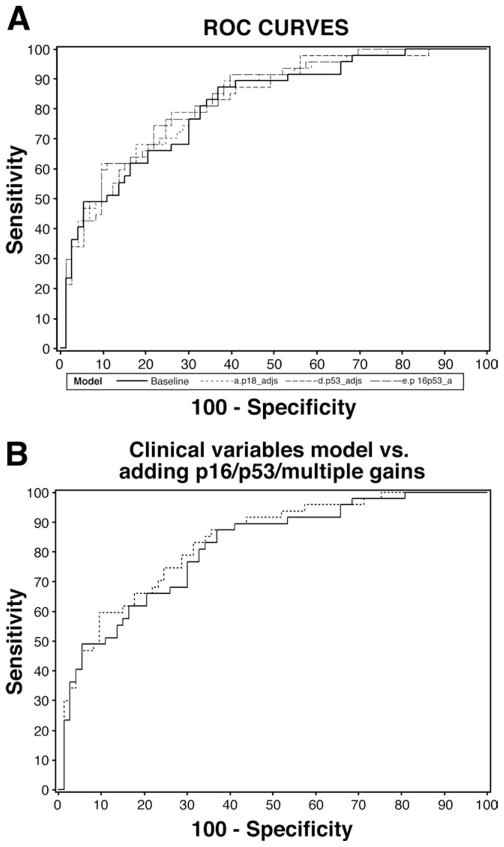
Results: A total of 126 consecutive patients (71 who underwent PDT and 55 patients who did not undergo PDT and were under surveillance, to adjust for the natural history of HGD), were included in this study. Fifty (40%) patients were responders (no dysplasia or carcinoma) at 3 months after PDT. On multiple variable analysis, P16 allelic loss (odds ratio [OR], 0.32; 95% confidence interval [CI], 0.10-0.96) predicted decreased response to PDT. BE segment length (OR, 0.71; 95% CI, 0.59-0.85), and performance of PDT (OR, 7.17; 95% CI, 2.50-20.53) were other independent predictors of loss of dysplasia.
Conclusions: p16 loss detected by fluorescence in situ hybridization can help predict loss of dysplasia in patients with BE and HGD/mucosal cancer. Biomarkers may help in the selection of appropriate therapy for patients and improve treatment outcomes.
Figures
Comment in
-
Biomarkers and photodynamic therapy for Barrett's esophagus: time to FISH or cut bait?Gastroenterology. 2008 Aug;135(2):354-7. doi: 10.1053/j.gastro.2008.06.065. Epub 2008 Jul 9. Gastroenterology. 2008. PMID: 18619447 No abstract available.
References
-
- Devesa SS, Blot WJ, Fraumeni JF., Jr Changing patterns in the incidence of esophageal and gastric carcinoma in the United States. Cancer. 1998;83:2049–2053. - PubMed
-
- Provenzale D, Schmitt C, Wong JB. Barrett’s esophagus: a new look at surveillance based on emerging estimates of cancer risk. Am J Gastroenterol. 1999;94:2043–2053. - PubMed
-
- Schnell TG, Sontag SJ, Chejfec G, et al. Long-term nonsurgical management of Barrett’s esophagus with high-grade dysplasia. Gastroenterology. 2001;120:1607–1619. - PubMed
-
- Weston AP, Sharma P, Topalovski M, et al. Long-term follow-up of Barrett’s high-grade dysplasia. Am J Gastroenterol. 2000;95:1888–1893. - PubMed
Publication types
MeSH terms
Substances
Grants and funding
LinkOut - more resources
Full Text Sources
Other Literature Sources
Medical
Research Materials
Miscellaneous
