. 2008 Jul;29(7):631-8.
doi: 10.1016/j.placenta.2008.04.007. Epub 2008 Jun 5.

Novel HLA-G-binding leukocyte immunoglobulin-like receptor (LILR) expression patterns in human placentas and umbilical cords

Affiliations

Novel HLA-G-binding leukocyte immunoglobulin-like receptor (LILR) expression patterns in human placentas and umbilical cords

R H McIntire et al. Placenta. 2008 Jul.

Abstract

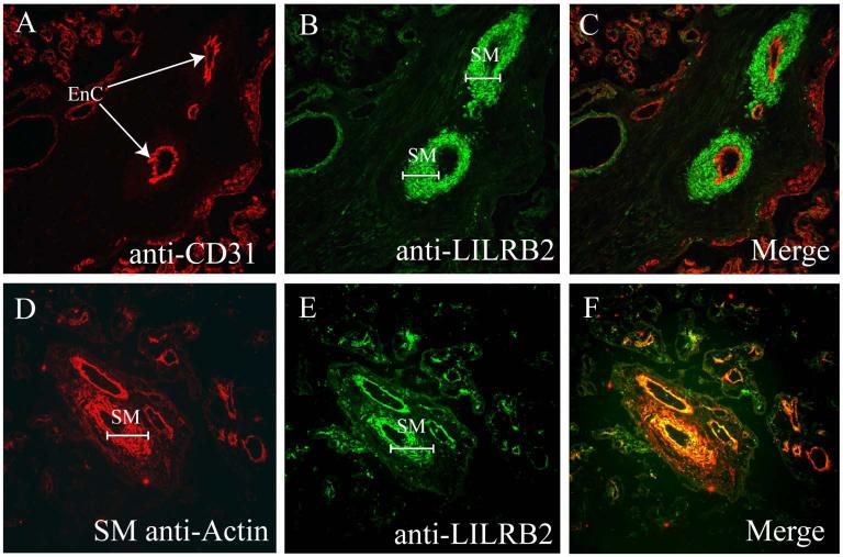
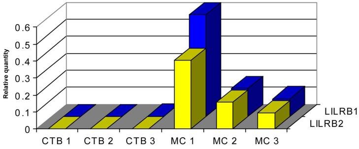
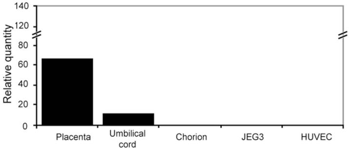
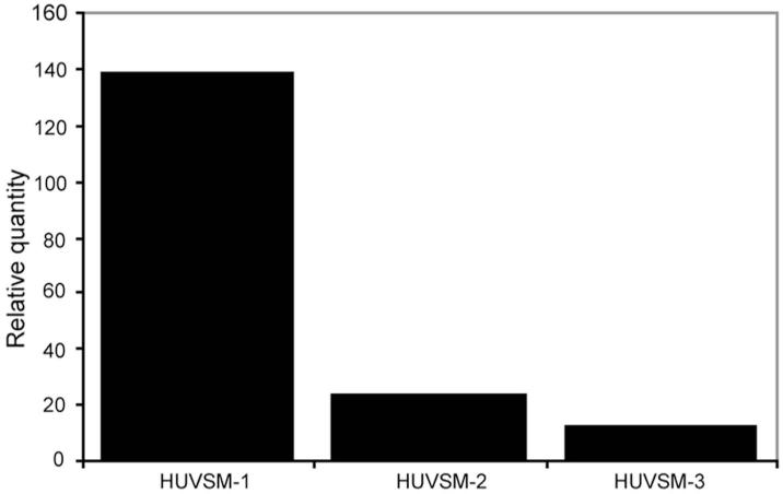
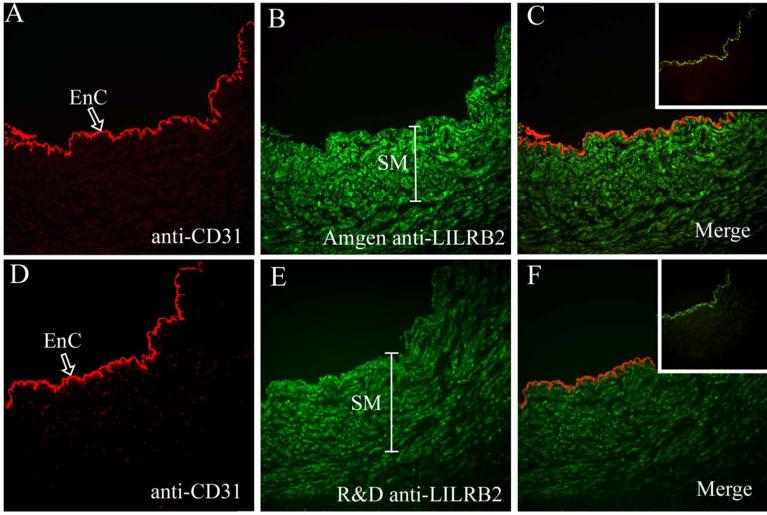
Human placentas are sources of cytokines, hormones and other substances that program receptive cells. One of these substances is HLA-G, which influences the functioning of both leukocytes and endothelial cells. In this study, we investigated the possibility that these and/or other types of cells in extraembryonic fetal tissues might respond to HLA-G by interacting with one or another of the leukocyte immunoglobulin-like receptors (LILR). LILRB1 is expressed by most leukocytes and LILRB2 is expressed primarily by monocytes, macrophages and dendritic cells. Analysis of term placentas by immunohistochemistry and Real Time PCR demonstrated that LILRB1 and LILRB2 protein and specific messages are produced in the mesenchyme of term villous placenta but are differently localized. LILRB1 was abundant in stromal cells and LILRB2 was prominent perivascularly. Neither receptor was identified in trophoblast. Further investigation using double label immunofluorescence indicated that placental vascular smooth muscle but not endothelia exhibit LILRB2. Term umbilical cord exhibited the same LILRB2 patterns as term placenta. Samples obtained by laser capture dissection of vascular smooth muscle in umbilical cords demonstrated LILRB2 mRNA, and double label immunofluorescence showed that cord vascular smooth muscle but not endothelium exhibited LILRB2 protein. The presence of LILRB1 in placental stromal cells and LILRB2 in vascular smooth muscle strongly suggest that HLA-G has novel functions in these tissues that could include regulation of placental immunity as well as development and function of the extraembryonic vasculature.


Figures

Fig. 1
Fig. 1. Identification of LILRB1 and LILRB2 proteins in human term villous placentas by immunohistology
(Upper left panel) Anti-LILRB1 (Amgen) fails to reveal signals in mesenchymal cells cuffing a small artery (large arrow) and a small vein (small arrow) but is strongly positive with pleiomorphic stromal cells (arrowhead). (Upper right panel) Anti-LILRB2 (Amgen) identifies positive cells cuffing a small artery (large arrow) and a small vein (small arrow) but does not yield readily identifiable signal in stromal cells (arrowhead). (Lower left panel) Anti-LILRB1 (Amgen) fails to reveal signals in cells cuffing a small vessel (large arrow) but is strongly positive with pleiomorphic stromal cells (arrowhead). (Lower right panel) Anti-LILRB2 (Amgen) identifies positive cells cuffing a small vessel (large filled arrow) and identifies diffuse, punctuate staining within the stroma (open arrows). Upper panels, original magnifications ×200; lower panels, original magnifications ×400. Positive stains in these experiments are identified by red signals. The slides were counterstained with Mayer’s hematoxylin.
Fig. 2
Fig. 2. Identification of LILRB2 positive vascular smooth muscle cells and LILRB2 negative endothelial cells in term villous placenta by double label immunofluorescence
(A) Anti-CD31. Arrows point to CD31+ arterial endothelial cells (EnC) in a term placental villus. (B) Anti-LILRB2. Brackets mark LILRB2+ smooth muscle (SM) cuffs around small arteries. (C) Merging of frames (A) and (B) reveals no double staining (yellow) in either endothelial cells or smooth muscle. (D) Anti-smooth muscle actin marks smooth muscle (SM) cuffs around villous placental vessels. (E) Anti-LILRB2. As in Panel (B), LILRB2 is prominent in smooth muscle (SM) cuffs around placental vessels. (F) Merging of frames (D) and (E) demonstrates bright yellow staining indicating that the vascular smooth muscle is LILRB2+. Images were captured at magnification ×200.
Fig. 3
Fig. 3. Use of Real Time PCR to identify LILRB1 and LILRB2 mRNA in highly purified term placental villous CTB cells and villous MC
Three preparations of purified CTB cells from three different term placentas failed to demonstrate LILRB1 (blue bars) or LILRB2 (yellow bars) mRNA. The matching villous MC from the same three placentas all demonstrated LILRB1 and LILRB2 mRNA. Levels of LILRB mRNA in placental cells were quantified against levels in PBMC.
Fig. 4
Fig. 4. Identification of LILRB2 mRNA in villous placenta, umbilical cord and cell lines by Real Time PCR
Levels of LILRB2 mRNA were assessed against levels of the mRNA in PBMC. (Lane 1) term villous placenta; (Lane 2) term umbilical cord; (Lane 3) chorion membrane CTB cells; (Lane 4) JEG-3 trophoblast tumor cell line; (Lane 5) human umbilical vein endothelial cells (HUVEC). Placenta and umbilical cord contained detectable levels of LILRB2 mRNA whereas chorion, JEG-3 and HUVEC cells did not contain the specific messages.
Fig. 5
Fig. 5. Use of Real Time PCR to identify LILRB2 mRNA in 3 different samples of umbilical cord vascular smooth muscle obtained by laser microdissection
Levels of LILRB2 mRNA in human umbilical vein smooth muscle (HUVSM) were compared with levels in PBMC to obtain relative quantity.
Fig. 6
Fig. 6. Double label immunofluorescence identification of CD31 and LILRB2 in umbilical cord vein
(A) Anti-CD31 recognizes venous endothelial cells (EnC, arrow). (B) Anti-LILRB2 (Amgen) identifies venous smooth muscle (SM, bracket), (C) Merging of the anti-CD31 and anti-LILRB2 (Amgen) demonstrates no bright yellow staining and therefore no overlap in expression of the two cellular markers. (D) Anti-CD31 recognizes venous endothelial cells, (EnC, arrow), (E) Anti-LILRB2 (R & D) identifies venous smooth muscle (SM, bracket), (F) Merging of the anti-CD31 and anti-LILRB2 (R & D) demonstrates no overlap in staining. Inserts to (C) and (F) are merged images of staining controls. Normal sheep Ig followed by the fluorochrome-labeled secondary antibody yielded no red signal but isotype-matched normal mouse Ig followed by the fluorochrome-labeled secondary antibody yielded a weak green signal on the inner elastic membrane located between the endothelium and smooth muscle layers. Original magnifications, ×200.

Comment in

References

    1. Ellis SA, Sargent IL, Redman CW, McMichael AJ. Evidence for a novel HLA antigen found on human extravillous trophoblast and a choriocarcinoma cell line. Immunol. 1986;59:595–601. - PMC - PubMed
    1. Kovats S, Main EK, Librach C, Stubblebine M, Fisher SJ, DeMars R. A class I antigen, HLA-G, expressed in human trophoblasts. Science. 1990;248:220–23. - PubMed
    1. Morales PJ, Pace JL, Platt JS, Phillips TA, Morgan K, Fazleabas AT, Hunt JS. Placental cell expression of HLA-G2 isoforms is limited to the invasive trophoblast phenotype. J Immunol. 2003;171:6215–24. - PubMed
    1. Morales PJ, Pace JL, Platt JS, Langat DL, Hunt JS. Synthesis of β2-microglobulin-free, disulfide-linked HLA-G5 homodimers in human placental villous cytotrophoblast cells. Immunol. 2007;122:179–88. 2007. - PMC - PubMed
    1. Hunt JS, Petroff MG, McIntire RH, Ober C. HLA-G and immune tolerance in pregnancy. FASEB J. 2005;19:681–93. - PubMed

Publication types

MeSH terms