Review
. 2008 Nov;22(6):321-32.
doi: 10.1016/j.blre.2008.04.003. Epub 2008 Jun 5.

Genetic causes of erythrocytosis and the oxygen-sensing pathway

Affiliations
Review

Genetic causes of erythrocytosis and the oxygen-sensing pathway

Frank S Lee. Blood Rev. 2008 Nov.

Abstract

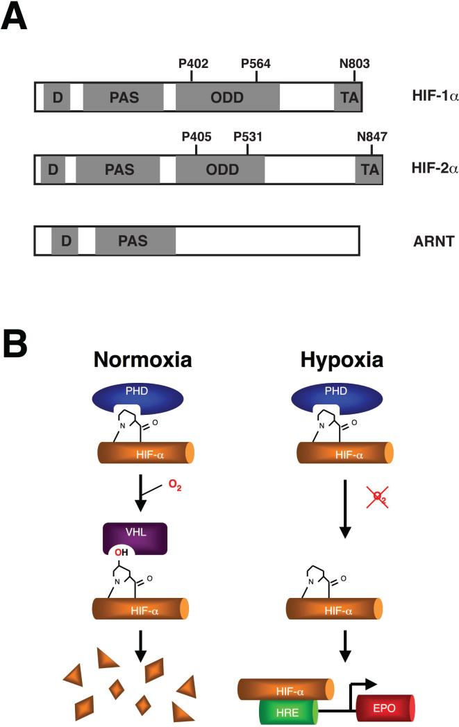
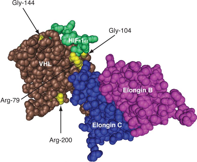
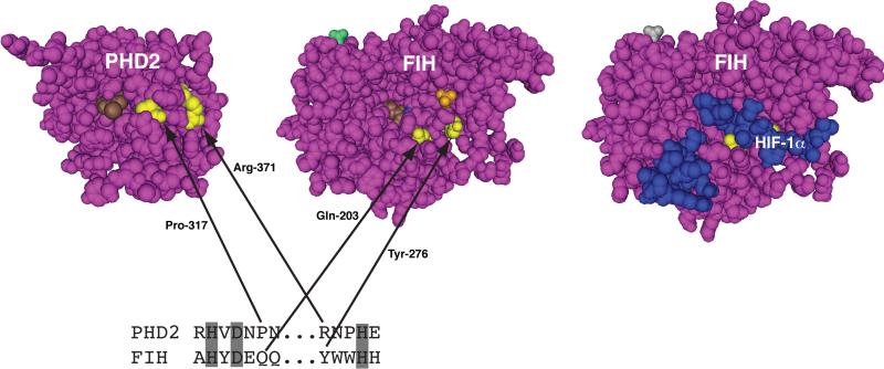
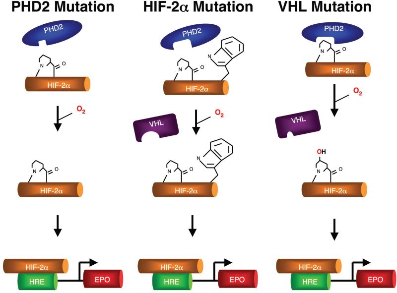
Idiopathic erythrocytosis is an uncommon disease, and is defined by an increase in red blood cell mass. The differential diagnosis of erythrocytosis is extensive, and can be divided into primary and secondary forms. Primary erythrocytoses are due to intrinsic defects in erythroid precursor cells and are characterized by low erythropoietin levels. Secondary erythrocytoses are extrinsic to erythroid progenitors and are characterized by either high or inappropriately normal erythropoietin levels. A distinct subset of secondary erythrocytoses are due to genetic mutations in key proteins of the oxygen-sensing pathway. These proteins constitute the core molecular machinery of oxygen-sensing with respect to red blood cell control. Apart from assigning physiologic roles for these proteins, studies of these rare mutations have (i) revealed the exquisite sensitivity of this pathway to genetic perturbations, (ii) highlighted important functional regions of the proteins, and (iii) provided a basis for potentially targeting this pathway for therapeutic benefit.


Figures

Figure 1
Figure 1
(A) Schematic diagrams of HIF-1α, HIF-2α, and ARNT. The DNA binding (D), Per-ARNT-Sim (PAS), oxygen-dependent degradation (ODD), and transcriptional activation (TA) domains are shaded and as indicated. The sites of prolyl (P402, P564, P405, P531) and asparaginyl hydroxylation (N803, N847) in HIF-1α and HIF-2α are indicated. (B) Regulation of HIF-α. Under normoxic conditions, HIF-α is prolyl hydroxylated, allowing binding of VHL which in turn promotes the degradation of HIF-α via ubiquitination and proteasomal degradation. Under hypoxic conditions, the prolyl hydroxylation is inhibited, thereby allowing stabilization of HIF-α and activation of HRE-driven genes, including the EPO gene.
Figure 2
Figure 2
Positions of select VHL-associated erythrocytosis mutations. Three-dimensional structure of the VCB complex bound to hydroxyproline-564 HIF-1α peptide (residues 556-575). The structure was generated using Cn3D from PDB coordinates (1LM8) deposited by Min et al. The positions of Arg-79, Gly-104, Gly-144, and Arg-200 in VHL are shown in yellow.
Figure 3
Figure 3
Positions of PHD2-associated erythrocytosis missense mutations. Three-dimensional structures of PHD2, FIH, and the FIH:HIF-1α (786-826) complex, generated using Cn3D from PDB coordinates (2G1M, 1H2N, and 1H2L) deposited by McDonough et al and Elkins et al . Arg-371 and Pro-317 in PHD2, and Tyr-276 and Gln-203 in FIH are highlighted in yellow. In the PHD2 structure, Compound A (a 2-oxoglutarate competitive inhibitor) is shown in brown. In the FIH structure, 2-oxoglutarate and sulfate are shown in brown and orange, respectively. In the FIH:HIF-1α (786-826) structure, the HIF-1α peptide is blue. Note that only HIF-1α residues 795-806 and 813-822 are resolved. At bottom is a comparison of residues 312-318 and 371-375 of PHD2 (separated by ...), and residues 198-204 and 276-280 of FIH. Shading indicates iron-chelating residues.
Figure 4
Figure 4
Three distinct types of mutations in the oxygen-sensing pathway can cause erythrocytosis. Mutations in PHD2 impair hydroxylation of HIF-2α (left), a mutation (Gly537Trp) in HIF-2α impairs PHD2-induced hydroxylation as well as subsequent recognition by VHL (middle), and mutations in VHL impair its capacity to induce HIF-2α degradation (right).

References

    1. Semenza GL. Life with oxygen. Science. 2007;318:62–4. - PubMed
    1. Wang GL, Jiang BH, Rue EA, Semenza GL. Hypoxia-inducible factor 1 is a basic-helix-loop-helix-PAS heterodimer regulated by cellular O2 tension. Proc Natl Acad Sci U S A. 1995;92:5510–4. - PMC - PubMed
    1. Gu YZ, Moran SM, Hogenesch JB, Wartman L, Bradfield CA. Molecular characterization and chromosomal localization of a third alpha-class hypoxia inducible factor subunit, HIF3alpha. Gene Expr. 1998;7:205–13. - PMC - PubMed
    1. Ema M, Taya S, Yokotani N, et al. A novel bHLH-PAS factor with close sequence similarity to hypoxia- inducible factor 1alpha regulates the VEGF expression and is potentially involved in lung and vascular development. Proc Natl Acad Sci U S A. 1997;94:4273–8. - PMC - PubMed
    1. Hogenesch JB, Chan WK, Jackiw VH, et al. Characterization of a subset of the basic-helix-loop-helix-PAS superfamily that interacts with components of the dioxin signaling pathway. J Biol Chem. 1997;272:8581–93. - PubMed

Publication types