. 2008 Jun 6;30(5):599-609.
doi: 10.1016/j.molcel.2008.03.020.

Distinct eRF3 requirements suggest alternate eRF1 conformations mediate peptide release during eukaryotic translation termination

Affiliations

Distinct eRF3 requirements suggest alternate eRF1 conformations mediate peptide release during eukaryotic translation termination

Hua Fan-Minogue et al. Mol Cell. .

Abstract

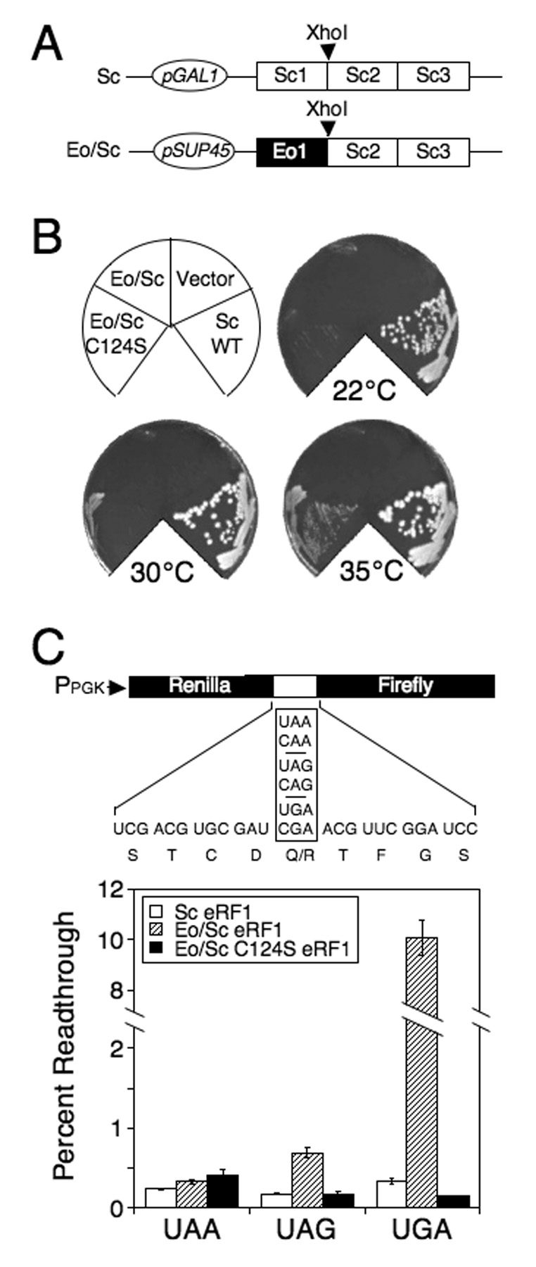
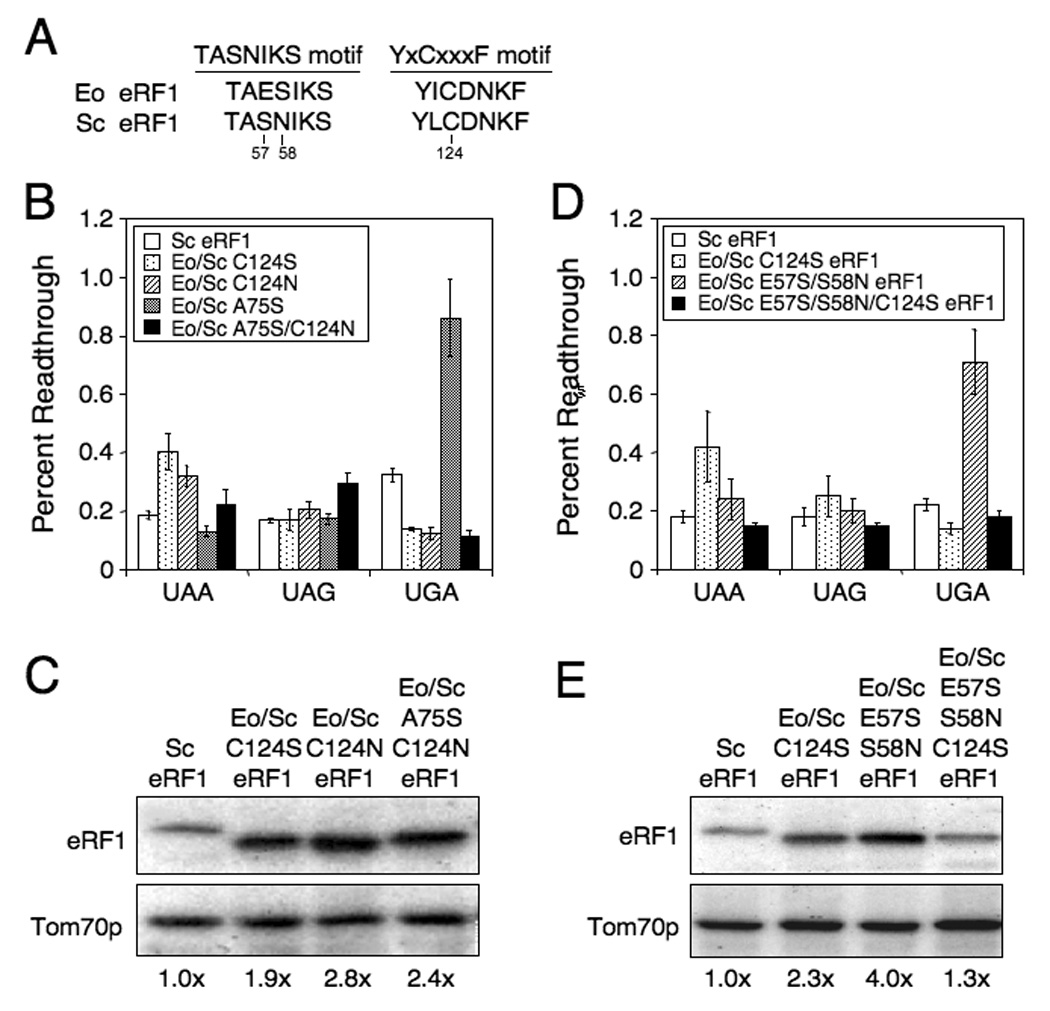
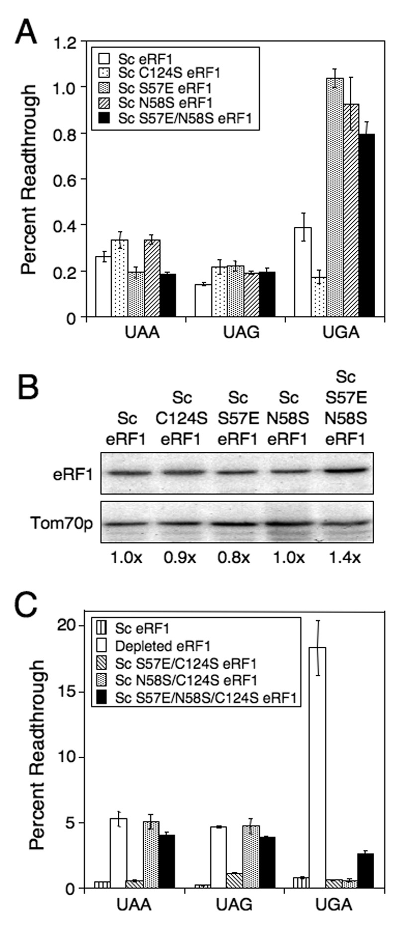
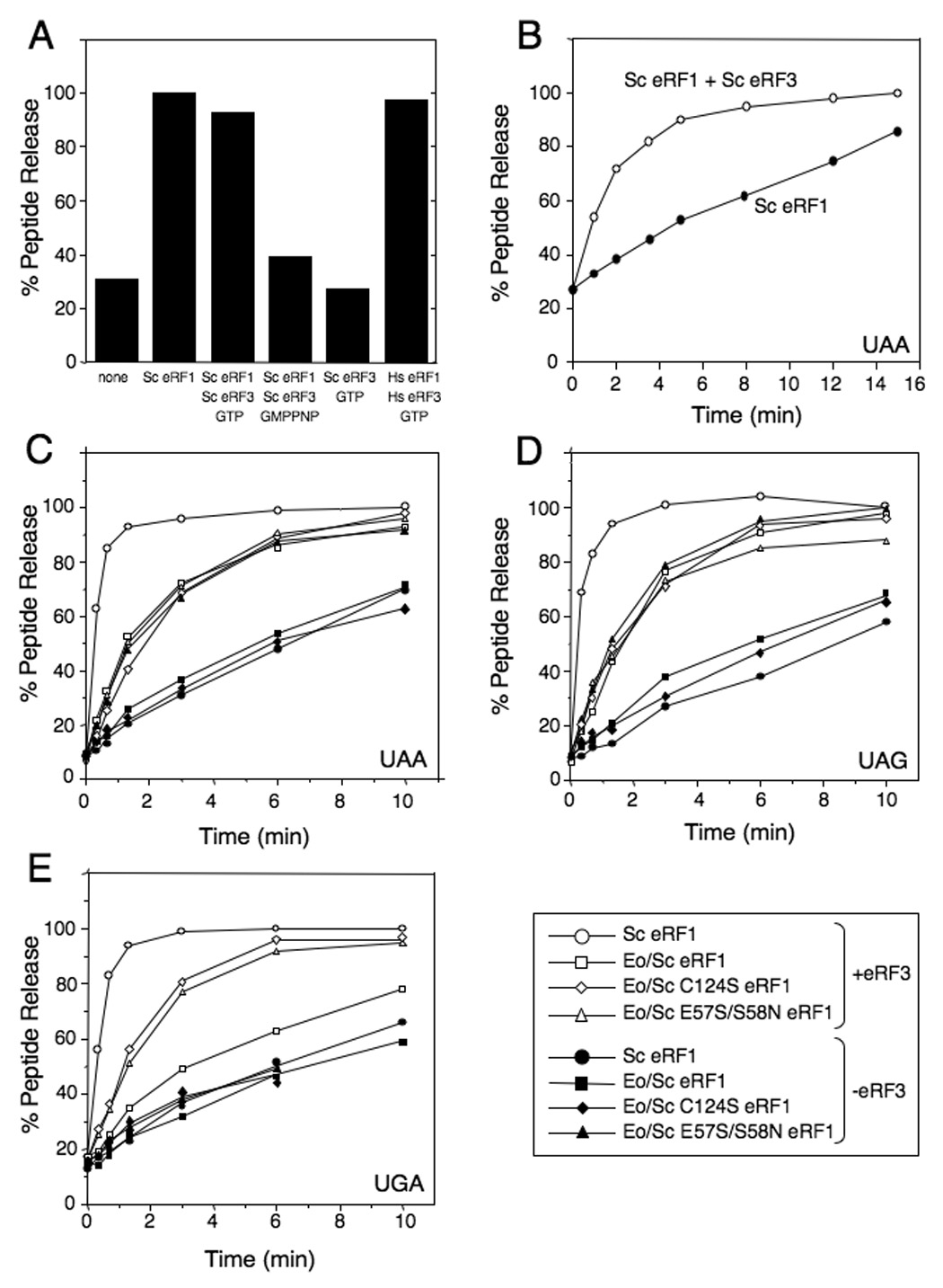
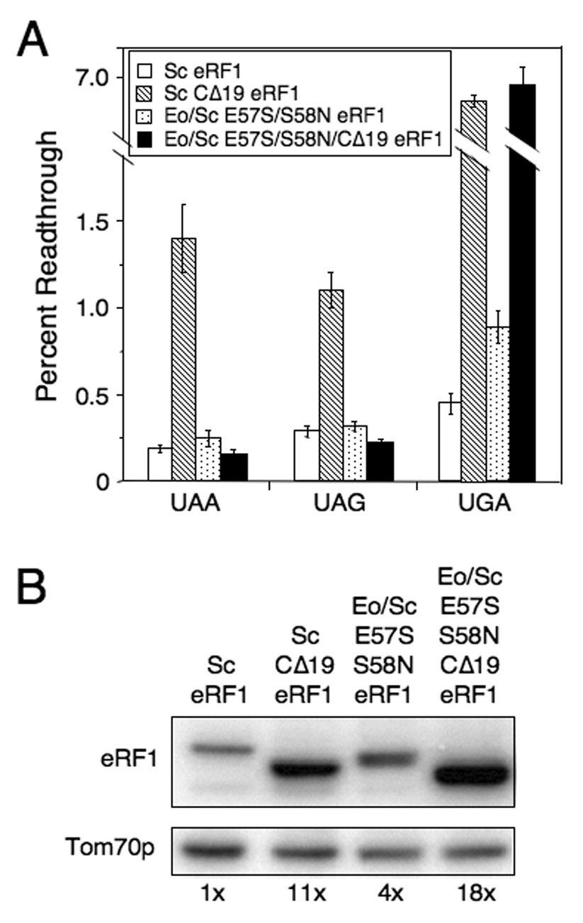
Organisms that use the standard genetic code recognize UAA, UAG, and UGA as stop codons, whereas variant code species frequently alter this pattern of stop codon recognition. We previously demonstrated that a hybrid eRF1 carrying the Euplotes octocarinatus domain 1 fused to Saccharomyces cerevisiae domains 2 and 3 (Eo/Sc eRF1) recognized UAA and UAG, but not UGA, as stop codons. In the current study, we identified mutations in Eo/Sc eRF1 that restore UGA recognition and define distinct roles for the TASNIKS and YxCxxxF motifs in eRF1 function. Mutations in or near the YxCxxxF motif support the cavity model for stop codon recognition by eRF1. Mutations in the TASNIKS motif eliminated the eRF3 requirement for peptide release at UAA and UAG codons, but not UGA codons. These results suggest that the TASNIKS motif and eRF3 function together to trigger eRF1 conformational changes that couple stop codon recognition and peptide release during eukaryotic translation termination.


Figures

Figure 1
Figure 1
Mutagenesis of Eo/Sc eRF1. (A) The Eo/Sc eRF1 plasmid used for random mutagenesis and the Sc eRF1 plasmid under GAL1 promoter control used to support growth of the sup45Δ strain while screening for suppressors of the hybrid Eo/Sc eRF1. (B) Growth of strain carrying Eo/Sc C124S eRF1. Plates were incubated at 22°C, 30°C and 35°C for 5 days. (C) Readthrough levels of wild type Sc eRF1, Eo/Sc eRF1 and Eo/Sc C124S eRF1 at 35°C. Readthrough in strains carrying the Eo/Sc eRF1 that cannot support cell growth as the sole source of eRF1 was carried out using a galactose to glucose shift protocol as described in the Materials and Methods. Readthrough values are represented as mean ± SD.
Figure 2
Figure 2
Efficiency of stop codon recognition mediated by Eo/Sc eRF1 suppressor mutants. (A) Alignment of TASNIKS and YxCxxxF motifs from Saccharomyces eRF1 and Euplotes eRF1. Numbering from Saccharomyces eRF1 is used. (B) Readthrough of stop codons measured in strains expressing wild type Sc eRF1 or Eo/Sc C124S, C124N, A75S or A75S/C124N eRF1. (C) Western blot quantitation of eRF1 levels from strains in (B). (D) Readthrough of stop codons measured in strains expressing wild type Sc eRF1 or Eo/Sc C124S, E57S/S58N, or E57S/S58N/C124S eRF1. (E) Western blot quantitation of eRF1 levels from strains in (D). All strains were grown in SM glucose medium at 35°C. Readthrough values are represented as mean ± SD.
Figure 3
Figure 3
Efficiency of stop codon recognition mediated by Sc eRF1 carrying C124S or TASNIKS mutations. (A) Readthrough of stop codons measured in strains expressing Sc eRF1 with C124S or viable TASNIKS mutations. (B) Western blot quantitation of eRF1 levels from strains in (A). (C) Readthrough in strains carrying Sc eRF1, depleted of eRF1, or carrying combinations of Sc eRF1 mutations that result in an inability to support growth as the sole source of eRF1. For mutant eRF1 proteins unable to support growth, readthrough was measured using a galactose to glucose shift protocol as described in the Materials and Methods. All strains were grown in SM glucose medium at 35°C. Readthrough values are represented as mean ± SD.
Figure 4
Figure 4
Eo/Sc eRF1 mutants restore peptide release at the UGA stop codon. A) Endpoint peptide release assay with excess Sc eRF1 and Sc eRF3. B) Kinetics of peptide release with Sc eRF1 (+/− Sc eRF3). C) Kinetics of peptide release with Eo/Sc eRF1 and mutant derivatives (+/− Sc eRF3) at UAA stop codon. D) Kinetics of peptide release with Eo/Sc eRF1 and mutant derivatives (+/− Sc eRF3) at UAG stop codon. E) Kinetics of peptide release with Eo/Sc eRF1 and mutant derivatives (+/− Sc eRF3) at UGA stop codon. Representative data for each experiment is shown. Reactions with eRF1s are denoted by closed symbols, while reactions with eRF1s and Sc eRF3 are denoted by open symbols.
Figure 5
Figure 5
Effect of reducing eRF3 binding on Eo/Sc E57S/S58N eRF1 function in vivo. A) Readthrough in strains carrying Sc eRF1, Sc CΔ19 eRF1, Eo/Sc E57S/S58N eRF1, or Eo/Sc E57S/S58N/CΔ19 eRF1. B) Western blot quantitation of eRF1 levels in strains above. All strains were grown in SM glucose medium at 30°C. Readthrough values are represented as mean ± SD.
Figure 6
Figure 6
Models of eukaryotic stop codon recognition and translation termination. A) The location of key determinants of eRF1 function on the three-dimensional structure of human eRF1. Only the relevant portion of domain 1 is shown. Residues identified by genetic screens in this study or by Stansfield and co-workers (Bertram et al., 2000) are indicated in yellow (residues identified in this study are in black type, while those identified by Stansfield and co-workers are in purple type). White circles indicate cavities 1, 2, and 3. The residue designations and numbering corresponds to human eRF1 (+3 relative to Sc eRF1). B) Model for eukaryotic translation termination at UAA/UAG vs. UGA stop codons.

References

    1. Alkalaeva EZ, Pisarev AV, Frolova LY, Kisselev LL, Pestova TV. In Vitro Reconstitution of Eukaryotic Translation Reveals Cooperativity between Release Factors eRF1 and eRF3. Cell. 2006;125:1125–1136. - PubMed
    1. Bertram G, Bell HA, Ritchie DW, Fullerton G, Stansfield I. Terminating eukaryote translation: domain 1 of release factor eRF1 functions in stop codon recognition. RNA. 2000;6:1236–1247. - PMC - PubMed
    1. Bonetti B, Fu L, Moon J, Bedwell DM. The efficiency of translation termination is determined by a synergistic interplay between upstream and downstream sequences in Saccharomyces cerevisiae. J Mol Biol. 1995;251:334–345. - PubMed
    1. Chavatte L, Seit-Nebi A, Dubovaya V, Favre A. The invariant uridine of stop codons contacts the conserved NIKSR loop of human eRF1 in the ribosome. EMBO J. 2002;21:5302–5311. - PMC - PubMed
    1. Eurwilaichitr L, Graves FM, Stansfield I, Tuite MF. The C-terminus of eRF1 defines a functionally important domain for translation termination in Saccharomyces cerevisiae. Mol Microbiol. 1999;32:485–496. - PubMed

Publication types

MeSH terms

LinkOut - more resources