. 2008 Aug 1;377(2):364-78.
doi: 10.1016/j.virol.2008.04.045. Epub 2008 Jun 9.

Relationship of HIV-1 and SIV envelope glycoprotein trimer occupation and neutralization

Affiliations

Relationship of HIV-1 and SIV envelope glycoprotein trimer occupation and neutralization

Emma T Crooks et al. Virology. .

Abstract

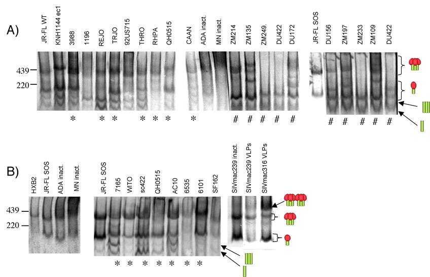
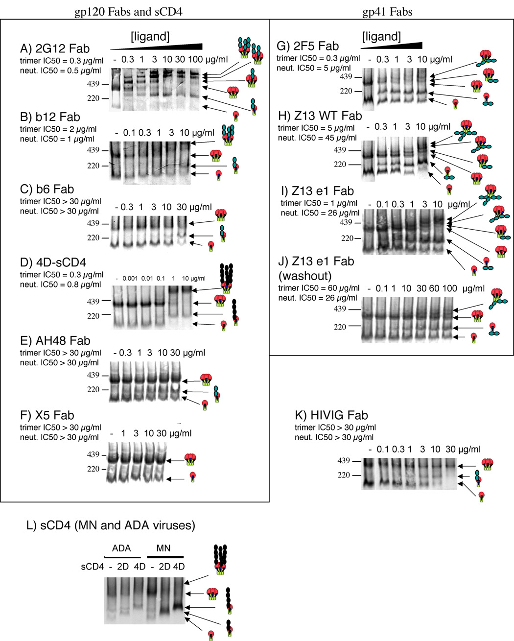
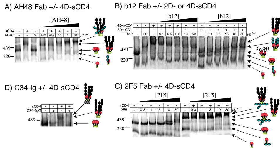
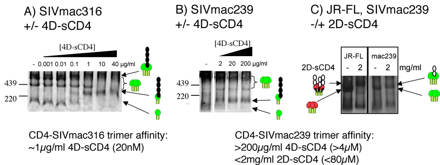
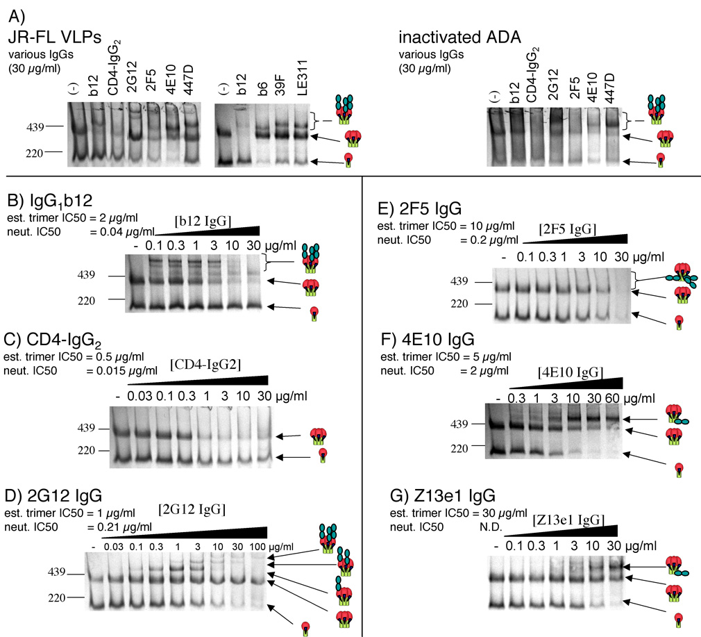
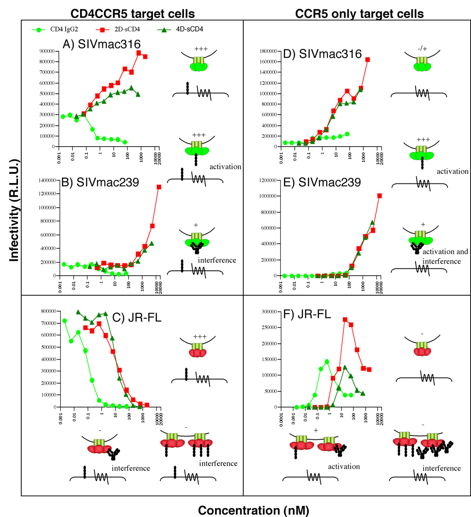
Insights into the process of HIV-1 neutralization may assist rational vaccine design. Here, we compared antibody neutralization against the JR-FL primary isolate and trimer binding affinities judged by native PAGE. Monovalent Fab-trimer binding and neutralization showed a direct quantitative relationship, implying that neutralization begins as each trimer is occupied by one antibody. At saturation, three Fab or soluble CD4 molecules engaged each trimer. In contrast, a maximum of one soluble CD4 molecule bound to functional SIV trimers with a truncated a gp41 tail. Remarkably, soluble CD4 was found to trigger dramatic enhancement of this virus. Unlike Fabs, a quantitative correlation between JR-FL trimer binding and neutralization was unclear for some, but not all IgGs, as neutralization was markedly increased, but trimer affinity was largely unchanged. In addition, only one molecule of certain gp41-specific IgGs appeared to be able to bind each trimer. We discuss the implications of these findings in weighing the relative contributions of size, multivalent binding and other possible effects of IgGs to explain their increased potency.


Figures

Fig. 1
Fig. 1. Behavior of native Env from various clade A, B and C HIV-1 viruses and SIVmac239 and SIVmac316 in native PAGE
Gp160ΔCT Env trimers derived from concentrated pseudovirion stocks were examined in BN-PAGE with reference to our JR-FL gp160ΔCT prototype. JR-FL gp160ΔCT trimers and monomers and gp41 trimers and monomers (Moore et al., 2006) are indicated by cartoons. In part A, and the middle section of part B, blots were probed with a cocktail of HIV+ donor plasmas. In the left and right panels of part B, blots were probed with either the HIV-1 gp120 mAb or SIV mAb cocktails, as described in materials and methods. Asterisks (*) denote Envs derived from a published clade B virus panel (Li et al., 2005) and a sharp sign (#) denotes Envs from a published clade C virus panel (Li et al., 2006).
Fig. 2
Fig. 2. Relationship of band density and gp120 quantity in BN-PAGE
Graded concentrations of JR-FL gp120 were loaded in BN-PAGE. Band densities were calculated and plotted against the microgram quantity of gp120.
Fig. 3
Fig. 3. Quantitative Ligand Binding to JR-FL trimers
Graded concentrations of Fabs or 4D-sCD4 were incubated with JR-FL SOS-VLPs Env trimers were then resolved by BN-PAGE. In one experiment (panel J), VLPs were washed to remove unbound Fab before native PAGE. Neutralizing IC50s and estimated trimer IC50s are indicated. Cartoons depict gp120/gp41 trimers and monomers and bound ligands. Black circles depict 4D-sCD4 and shaded circles depict Fab. The molecular weights of trimers and monomers were previously estimated as 420 kDa and 140 kDa (Moore et al., 2006). The masses of trimers complexed with saturating concentrations of neutralizing Fabs were estimated as follows: 2G12 (780 kDa), b12 (614 kDa), 4D-sCD4 (620 kDa), 2F5 (493 kDa), Z13 WT (551 kDa) and Z13e1 (533 kDa). In the case of the gp41 Fabs, the complexes with three Fabs were unclear, so the molecular weights given are estimated of trimers with two Fabs bound. Panel L shows the effects of saturating (20µg/ml) 2D-and 4D-sCD4 on live inactivated MN and ADA preparations.
Fig. 4
Fig. 4. Ligand binding to JR-FL in the presence of sCD4
The binding of various ligands to JR-FL SOS-VLP trimers was determined in the presence or absence of 2D- or 4D-sCD4 (5 µg/ml). A) Fab AH48. B) Fab b12. C) Fab 2F5. D) C34-Ig. In the case of 2F5, unbound Fab was washed away prior to sample preparation, but in all other cases, ligand was left in. A final concentration of 40 µg/ml (666 nM) C34-Ig was added to VLPs. Cartoons indicate liganded and unliganded trimers. Black circles depict 4D-sCD4, white circles depict 2D-sCD4, shaded circles depict Fab and C34-Ig. In part A, the size of the 4D-sCD4+AH48 saturated trimer (far right lane) was estimated as 780 kDa. In part C, the size of trimer+sCD4+2F5 complexes at near saturation (right lane) was estimated as 795 kDa. In part D, the sizes of complexed trimers were estimated as: lane 2 (trimer+C34-Ig) 447 kDa; lane 3 (trimer+4D-sCD4) 620 kDa; lane 4 (trimer+4D-sCD4+C34-Ig) 672 kDa.
Fig. 5
Fig. 5. Soluble CD4 affinity and stoichiometry for SIV trimers
Graded concentrations of 4D-sCD4 were mixed with A) SIVmac316 or B) SIVmac239 Env trimers in BN-PAGE. C) Saturating concentrations (2 mg/ml; 80 µM) of 2D-sCD4 were mixed with JR-FL and SIVmac239 Env trimers in BN-PAGE. Cartoons indicate bound and unbound forms of Env.
Fig. 6
Fig. 6. Binding of whole IgG mAbs to JR-FL trimers
A) Various IgGs (at 30 µg/ml) were incubated with JR-FL SOS-VLPs or ADA inactivated particles and resolved by BN-PAGE. B)-G) Increasing concentrations of IgG1b12, CD4-IgG2, 2G12, 2F5, 4E10, or Z13e1 were mixed with JR-FL SOS-VLPs. Unbound IgG was removed by a wash with PBS. Cartoons indicate liganded and unliganded trimers. Though the ligands are in the form of IgGs, for convenience, they are depicted as two shaded circles. N.D. = not done. Neutralizing IC50s and estimated trimer IC50s are indicated, where available. The trimer complexes with one 2G12 IgG molecule (part D, lane 5) were estimated at 696 kDa, those of 4E10 IgG and Z13e1 IgG were estimated as 626 kDa and 720 kDa, respectively.
Fig. 7
Fig. 7. Effect of CD4 binding stoichiometry on HIV-1 and SIV infection
2D-sCD4, 4D-sCD4 and CD4-IgG2 were incubated with SIVmac316 (A, D), SIVmac239 (B, E) or JR-FL (C, F) gp160ΔCT viruses, and infection in relative light units (R.L.U.) was measured on CF2 target cells bearing both CD4 and CCR5 (left panels) or CCR5 only (right panels). Baseline infection of CCR5-expressing targets with no sCD4 (not shown) was identical to that at the lowest sCD4 concentrations shown. The effects of CD4-based ligands on SIV and HIV-1 infection are illustrated by cartoons. The levels of infection in each cartoon scenario is depicted by + signs. Soluble CD4 (2D- or 4D-) is depicted as 4 black circles, and CD4-IgG2 is depicted as a series of black circles in an IgG conformation.

References

    1. Arthos J, Cicala C, Steenbeke TD, Chun TW, Dela Cruz C, Hanback DB, Khazanie P, Nam D, Schuck P, Selig SM, Van Ryk D, Chaikin MA, Fauci AS. Biochemical and biological characterization of a dodecameric CD4-Ig fusion protein: implications for therapeutic and vaccine strategies. J Biol Chem. 2002;277(13):11456–11464. - PubMed
    1. Barbas CF, 3rd, Collet TA, Amberg W, Roben P, Binley JM, Hoekstra D, Cababa D, Jones TM, Williamson RA, Pilkington GR, et al. Molecular profile of an antibody response to HIV-1 as probed by combinatorial libraries. J Mol Biol. 1993;230(3):812–823. - PubMed
    1. Bennett A, Liu J, Van Ryk D, Bliss D, Arthos J, Henderson RM, Subramaniam S. Cryoelectron tomographic analysis of an HIV-neutralizing protein and its complex with native viral gp120. J Biol Chem. 2007;282(38):27754–27759. - PubMed
    1. Binley JM, Cayanan CS, Wiley C, Schulke N, Olson WC, Burton DR. Redox-triggered infection by disulfide-shackled human immunodeficiency virus type 1 pseudovirions. J Virol. 2003;77(10):5678–5684. - PMC - PubMed
    1. Binley JM, Ditzel HJ, Barbas CF, 3rd, Sullivan N, Sodroski J, Parren PW, Burton DR. Human antibody responses to HIV type 1 glycoprotein 41 cloned in phage display libraries suggest three major epitopes are recognized and give evidence for conserved antibody motifs in antigen binding. AIDS Res Hum Retroviruses. 1996;12(10):911–924. - PubMed

Publication types

MeSH terms