. 2008 Sep;31(2):98-109.
doi: 10.1016/j.jaut.2008.04.022. Epub 2008 Jun 9.

Autoantibody production in lpr/lpr gld/gld mice reflects accumulation of CD4+ effector cells that are resistant to regulatory T cell activity

Affiliations

Autoantibody production in lpr/lpr gld/gld mice reflects accumulation of CD4+ effector cells that are resistant to regulatory T cell activity

Brian D Hondowicz et al. J Autoimmun. 2008 Sep.

Abstract

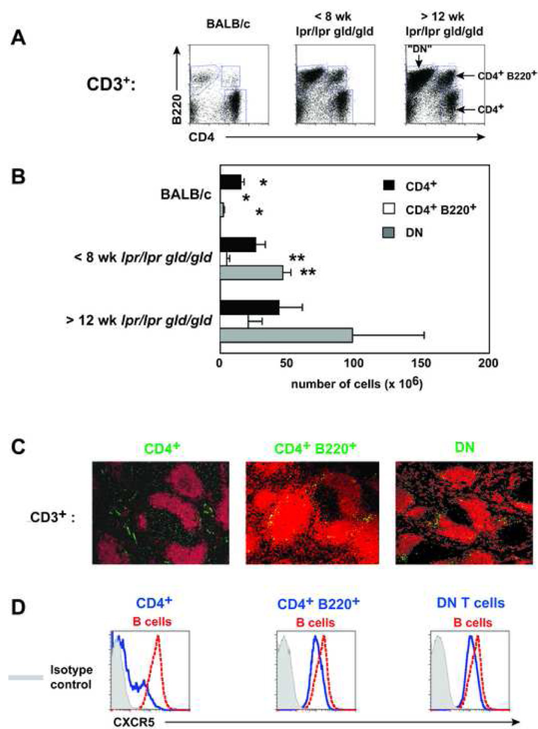
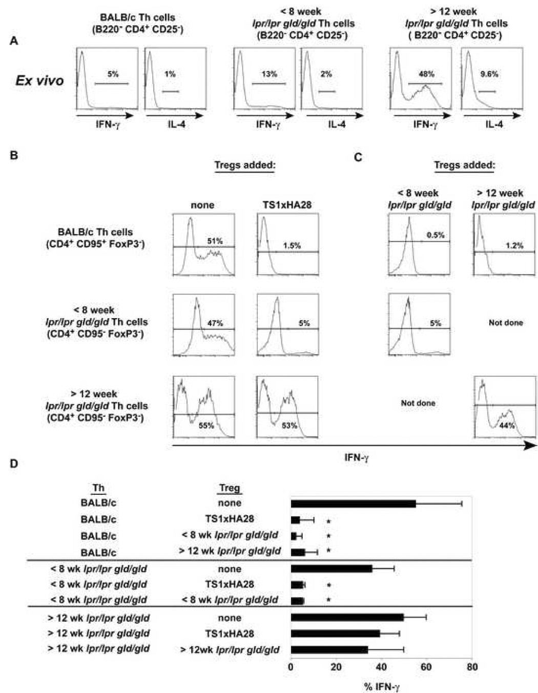
In Fas/FasL-deficient mice anti-chromatin Ab production is T cell dependent and is not apparent until after 10 weeks of age. Early control of anti-chromatin antibodies may be due to the counterbalancing influence of Treg cells. Here we show that Treg cells block lpr/lpr gld/gld Th cells from providing help to anti-chromatin B cells in an in vivo transfer system. Interestingly, the percentage and absolute numbers of Foxp3+ Treg cells is elevated in BALB/c-lpr/lpr gld/gld mice and increases with age compared to BALB/c mice. The majority of Foxp3 expression is found in the B220- CD4+ T cell population, and Foxp3-expressing cells are localized in the splenic PALS (periarteriolar lymphocyte sheath). Strikingly, although the lack of functional Fas/FasL does not affect the ability of Treg cells to block Th cell proliferation, Treg cells can block the IFN-gamma differentiation of Th cells from BALB/c or young BALB-lpr/lpr gld/gld mice but not of pre-existing Th1 cells from older BALB/c-lpr/lpr gld/gld mice. Thus, we suggest autoantibody production is not caused by the lack of Treg cells but by a defect in activation-induced cell death that leads to the accumulation of T effector cells that are resistant to regulatory T cell activity.


Figures

Figure 1
Figure 1. lpr/lpr gld/gld anti-chromatin B cells do produce autoAbs in the presence of Th cells
A) 20 ×106 TS1 lymph node cells were transferred into 4–5 week old VH3H9/HACII lpr/lpr gld/gld mice for 10 days. At day 10, B) serum or C) spleen was taken and assayed for ANAs and splenic localization, respectively (n=3).
Figure 2
Figure 2. Treg cells block in vivo autoAb production promoted by lpr/lpr gld/gld Th cells
A) In vivo protocol for determining whether young lpr/lpr gld/gld Th cells can be blocked from inducing autoreactive anti-chromatin antibody production. B) Anti-chromatin Abs from the serum of day 8 CB17 mice that received B cells alone (black circles), TS1 Th cells (black diamonds), TS1 Th + Treg cells (white diamonds), TS1 lpr/lpr gld/gld Th (black squares), and TS1 lpr/lpr gld/gld Th + Treg cells (white squares). Data are the average of three experiments. (*) indicates p < 0.05 and is different compared to all groups. C) Localization of IgMa cells in the spleen of the indicated groups by immunohistochemistry (n=3).
Figure 3
Figure 3. T cell subsets in the lymph node and spleen
A) Representative plots of CD3+ cells from the spleens of BALB/c and young and old lpr/lpr gld/gld mice (n=4). B) Total cell numbers from the spleen of BALB/c and lpr/lpr gld/gld mice (n=4). (*) indicates significantly different compared to all group of mice except for those labeled. (**) indicateds are significantly different than old lpr/lpr gld/gld mice only. C) The indicated T cell populations were sorted from >12 week old lpr/lpr gld/gld spleens, CFSE labeled, and transferred into BALB/c mice overnight. Recipient mice were stained with anti-B220 (red) and transferred cells were visualized by immunofluorescence (n=3). D) Histograms of CXCR5 expression on the T cell population indicated in C) (n=3).
Figure 4
Figure 4. Foxp3 expression in lpr/lpr gld/gld mice
A) Representative plots of Foxp3 in the spleens of the indicated T cell population. The average percentage of Foxp3+ cells in BALB/c mice: CD4+, inguinal LN = 10.9% +/− 2.3%, spleen = 6.8% +/− 2.6% (n=4); CD4+ B220+, inguinal LN = 12.0% +/− 3.0%, spleen = 12.1% +/− 1.7% (n=3). The average percentage of Foxp3+ cells in young lpr/lpr gld/gld mice: CD4+, inguinal LN = 11.9% +/− 6.4%, spleen = 18.0% +/− 5.0% (n=3–4); CD4+ B220+, inguinal LN = 4.0% +/− 1.6%, spleen = 3.9% +/− 0.8% (n=3). The average percentage of Foxp3+ cells in old lpr/lpr gld/gld mice: CD4+, inguinal LN = 14.7% +/− 3.7%, spleen = 25.8% +/− 5.0% (n=3–4); CD4+ B220+, inguinal LN = 1.1% +/− 0.5%, spleen = 2.5% +/− 0.7% (n=2–3). B) Absolute cell number of Foxp3+ and Foxp3 cells in the CD4+, CD4+ B220+, and DN T cell subsets (n=3). C) Foxp3 localization in the spleens of BALB/c and young and old lpr/lpr gld/gld mice (n≥3).
Figure 5
Figure 5. Treg and Th cells from lpr/lpr gld/gld mice can inhibit proliferation and be inhibited in vitro
A) 5×10⁴ Treg cells (CD4+ CD25+) from TS1xHA28 BALB/c mice were cultured with 5×10⁴ Th cells from young or old lpr/lpr gld/gld mice and stimulated as in B). The data is the average of three separate experiments. A) 5×10⁴ Treg (B220 CD4+ CD25+) cells from young and old lpr/lpr gld/gld mice were cultured with 5×10⁴ Th cells from BALB/c mice, 5×10⁵ irradiated BALB/c spleen cells and stimulated with anti-CD3. After 72h stimulation the cultures were pulsed with 3H thymidine and then harvested 16h later. The data is the average of three separate experiments.
Figure 6
Figure 6. The effect of Treg cells on the IFN-γ potential of Th cells
A) Th cells from the indicated mice were stimulated with PMA and Ionomycin ex vivo and 5 hours later were stained for IFN-γ and IL-4 (n=2). B) and C) Th cells and Treg cells were sorted from BALB/c and lpr/lpr gld/gld mice and stimulated with anti-CD3 for three days in the presence or absence of different Treg cell populations. On day 3 the cultures were stimulated with PMA and Ionomycin and stained for IFN-γ. The representative histograms are gated on CD4+ Foxp3 cells and the percentage of IFN-γ+ cells are shown (n=3). D) The bar graph shows the average frequency of cells making IFN-γ from all three experiments. * denotes p < 0.05 between Th cells with and without Treg cells.
Figure 7
Figure 7. The response of Th1 and Th2 cells to Treg cells
Using BALB/c TS1 TCR Tg mice, naïve, Th1, or Th2 cells were stimulated as in Figure 6 and stained for the number of cells producing IFN-γ or IL-4 (n=2).
Figure 8
Figure 8. Anti-chromatin Ab responses from anti-CD25 depleted gld/gld or lpr/lpr gld/gld mice
Weekly at 2–4 weeks of life 1 mg of PC61 (anti-CD25) was administered i.p. to A) gld/gld (n=3) or B) lpr/lpr gld/gld (n=2). Simultaneously control gld/gld or lpr/lpr gld/gld mice were treated with A) PBS (n=5) or B) Rat IgG (n=4), respectively. At weeks A) 5 and 6 or B) week 6, mice were bled and the isotype of anti-chromatin antibodies were determined. (*) denotes p<0.05 between PC61-treated and control treated mice.

References

    1. Mandik-Nayak L, Bui A, Noorchashm H, Eaton A, Erikson J. Regulation of anti-double-stranded DNA B cells in nonautoimmune mice: localization to the T-B interface of the splenic follicle. Journal of Experimental Medicine. 1997;186:1257–1267. - PMC - PubMed
    1. Mandik-Nayak L, Seo S-j, Sokol C, Potts KM, Bui A, Erikson J. MRL-lpr/lpr mice exhibit a defect in maintaining developmental arrest and follicular exclusion of anti-double-stranded DNA B cells. Journal of Experimental Medicine. 1999;189:1799–1814. - PMC - PubMed
    1. Fields ML, Sokol CL, Eaton-Bassiri A, Seo S-j, Madaio MP, Erikson J. Fas/Fas Ligand deficiency results in altered localization of anti-double-stranded DNA B cells and dendritic cells. Journal of Immunology. 2001;167:2370–2378. - PubMed
    1. Seo S-j, Fields ML, Buckler JL, Reed AJ, Mandik-Nayak L, Nish SA, Noelle RJ, Turka LA, Finkelman FD, Caton AJ, Erikson J. The impact of T helper and T regulatory cells on the regulation of anti-double-stranded DNA B cells. Immunity. 2002;16:535–546. - PubMed
    1. Fields ML, Hondowicz BD, Metzgar MH, Nish SA, Wharton GN, Picca CC, Caton AJ, Erikson J. CD4+CD25+ Regulatory T Cells Inhibit the Maturation but Not the Initiation of an Autoantibody Response. J Immunol. 2005;175:4255–4264. - PubMed

Publication types