

doi: 10.1016/j.tube.2008.04.002.
Epub 2008 Jun 9.
The immunomodulatory lipoglycans, lipoarabinomannan and lipomannan, are exposed at the mycobacterial cell surface
Affiliations
- PMID: 18539533
- PMCID: PMC2613510
- DOI: 10.1016/j.tube.2008.04.002
Item in Clipboard
The immunomodulatory lipoglycans, lipoarabinomannan and lipomannan, are exposed at the mycobacterial cell surface
Tuberculosis (Edinb).
2008 Nov.
Abstract
By labeling surface carbohydrates, we found that a pool of lipoglycans, cell wall associated, is exposed at the cell surface of mycobacteria and thus, most probably, inserted in the outer leaflet of the outer membrane. In contrast, plasma membrane anchored lipoglycans are not accessible to surface labeling. This result supports the role of lipoglycans as key immunomodulatory molecules but raises the question of their transport from the plasma membrane, where they are synthesized, to the outermost layers of the envelope, where they can act as modulins. The data are discussed in terms of consequences for cell envelope organization.
Figures

A) 1 μg of each fraction (10 μg for arabinogalactan) were dot-blotted and probed with AP-streptavidin. 1, control cells; 2, biotinylated cells. HIC, hydrophobic interaction chromatography. B) Bacteria were fixed with 2% glutaraldehyde (EMS, Washington PA) in 0.1 M cacodylate buffer pH 7.4 during 1 hour at 4°C. Fixed bacteria were washed in 0.2 M cacodylate buffer (pH 7.4), postfixed with 1% osmium tetroxide in 0.1 M cacodylate buffer for 1 h and dehydrated in graded ethanol series. After dehydration samples were critical point dried with an emscope CPD 750 apparatus, mounted on stubs, coated with gold-palladium alloy with a JEOL JFC 1100 ion sputtering apparatus and examined with a Hitachi S-450 scanning electron microscope at an accelerating voltage of 15 kV. Bars, 0.5 μm.

The cell wall (cw) and plasma membrane (pm) fractions were prepared from M. smegmatis cells as described in A) and submitted to α-amylase and protease digestions. Lipoglycans were recovered after HIC and LAM (5μg) was analyzed by: B) SDS-PAGE revealed by periodic acid-silver nitrate staining; C) western blot probed with AP-streptavidin; D) western blot probed with anti-LAM CS35 antibody. 1, LAM from non-labeled control cells; 2, LAM from biotin-hydrazide labeled cells; Sc, in vitro biotinylated M. smegmatis total LAM standard; Sd, M. smegmatis total LAM standard.
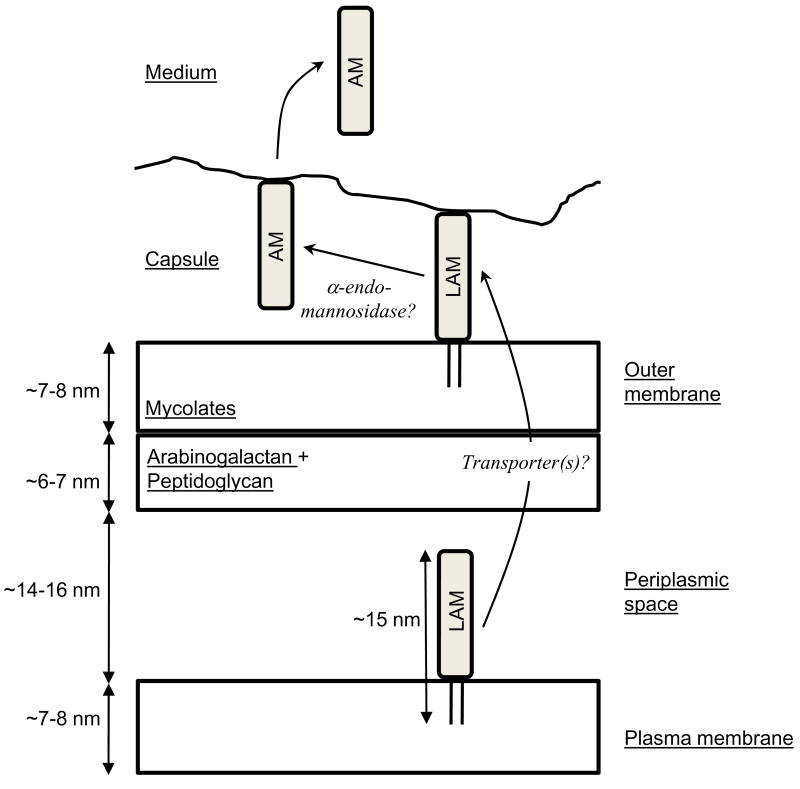
All the constituents are tentatively presented to scale and the drawing is based on the recent electron microscopy analyses on unperturbed cells ,. Lipoglycans are synthesized at the level of the plasma membrane where they have not access to cell surface. The hydrophilic part of the molecule is most probably located in the periplasmic space, which has now been clearly evidenced ,, or may protrude through the cell wall skeleton via the pores made by cross-linked peptidoglycan strands . After synthesis, a part of or all the lipoglycans are transferred, possibly via a transporter(s) that still remains to be discovered, to the outer layers of the cell envelope. At this stage, they are exposed at the cell surface and most probably inserted among other lipids in the outer layer of the outer membrane and can play their roles of modulins and adhesins. Action of an endogenous α-endomannosidase might convert a portion of lipoarabinomannan (LAM) and lipomannan molecules into their lipid free glycan counterparts, arabinomannan (AM) and mannan respectively that can be subsequently released into the culture medium.
References
-
- Chatterjee D, Khoo KH. Mycobacterial lipoarabinomannan: an extraordinary lipoheteroglycan with profound physiological effects. Glycobiology. 1998;8:113–20. - PubMed
-
- Nigou J, Gilleron M, Puzo G. Lipoarabinomannans: from structure to biosynthesis. Biochimie. 2003;85:153–66. - PubMed
-
- Briken V, Porcelli SA, Besra GS, Kremer L. Mycobacterial lipoarabinomannan and related lipoglycans: from biogenesis to modulation of the immune response. Mol Microbiol. 2004;53:391–403. - PubMed
-
- Sutcliffe I. Lipoarabinomannans--structurally diverse and functionally enigmatic macroamphiphiles of mycobacteria and related actinomycetes. Tuberculosis (Edinb) 2005;85:205–6. - PubMed
-
- Gilleron M, Jackson M, Nigou J, Puzo G. Structure, biosynthesis, and activities of the phosphatidyl-myo-inositol-based lipoglycans. In: Daffé M, Reyrat JM, editors. The Mycobacterial Cell Envelope. Washington, DC: ASM Press; 2008. pp. 75–105.
Publication types
MeSH terms
Substances
Grants and funding
LinkOut - more resources
Full Text Sources
