. 2008 Aug 1;283(31):21453-61.
doi: 10.1074/jbc.M800886200. Epub 2008 Jun 6.

The cytoplasmic domain of the hyaluronan receptor for endocytosis (HARE) contains multiple endocytic motifs targeting coated pit-mediated internalization

Affiliations

The cytoplasmic domain of the hyaluronan receptor for endocytosis (HARE) contains multiple endocytic motifs targeting coated pit-mediated internalization

Madhu S Pandey et al. J Biol Chem. .

Abstract

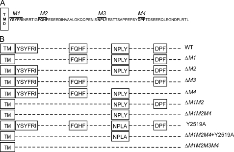
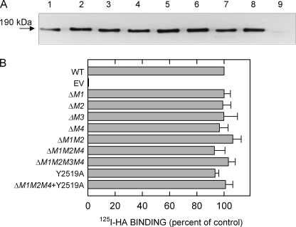
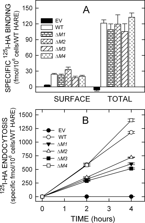
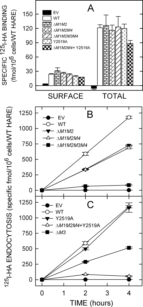
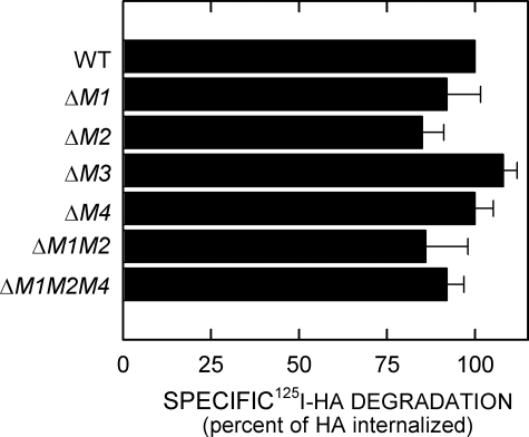
The hyaluronic acid (HA) receptor for endocytosis (HARE) is the primary scavenger receptor for HA and chondroitin sulfates in mammals. The two human isoforms of HARE (full-length 315-kDa and a 190-kDa proteolytic cleavage product), which are type I single-pass membrane proteins, are highly expressed in sinusoidal endothelial cells of lymph nodes, liver, and spleen. Their identical HARE cytoplasmic domains contain four candidate AP-2/clathrin-mediated endocytic signaling motifs as follows: YSYFRI(2485), FQHF(2495), NPLY(2519), and DPF(2534) (315-HARE numbering). Stably transfected cells expressing 190-HARE(DeltaYSYFRI), 190-HARE(DeltaFQHF), or 190-HARE(DeltaNPLY) (lacking Motifs 1, 2, or 3) had decreased (125)I-HA endocytosis rates of approximately 49, approximately 39, and approximately 56%, respectively (relative to wild type). In contrast, 190-HARE(DeltaDPF) cells (lacking Motif 4) showed no change in HA endocytic rate. Deletions of motifs 1 and 2 or of 1, 2, and 4 decreased the rate of HA endocytosis by only approximately 41%. Endocytosis was approximately 95% decreased in mutants lacking all four motifs. Cells expressing a 190-HARE(Y2519A) mutant of the NPLY motif retained 85-90% of wild type endocytosis, whereas this mutation in the triple motif deletant decreased endocytosis to approximately 7% of wild type. Tyr in NPLY(2519) is thus important for endocytosis. All HARE mutants showed similar HA binding and degradation of the internalized HA, indicating that altering endocytic motifs did not affect ectodomain binding of HA or targeting of internalized HA to lysosomes. We conclude that, although NPLY may be the most important motif, it functions together with two other endocytic motifs; thus three signal sequences (YSYFRI, FQHF, and NPLY) provide redundancy to mediate coated pit targeting and endocytosis of HARE.


Figures

FIGURE 1.
FIGURE 1.
The 190- and 315-HARE CD and mutants used in this study. A, four putative endocytic motifs (indicated in boldface and overlined) are present in the 72-aa CD of recombinant 190-HARE (1416 aa long) or 315-HARE (2551 aa long). The four sequence motifs (YSYFRI2485, FQHF2495, NPLY2519, and DPF2534) are referred to as M1, M2, M3, and M4, respectively. Numbering is relative to the full-length 315-HARE protein (13, 15). V5 and His6 epitopes are at the COOH termini of all 190-HARE mutants. B, scheme illustrates the various motif deletion or site-specific 190-HARE CD mutants used. Constructs and stably transfected Flp-In-293 cell lines were prepared as described under “Experimental Procedures.” TM, transmembrane.
FIGURE 2.
FIGURE 2.
Expression and 125I-HA ligand binding of 190-HARE CD mutants. Cells were grown to confluence, washed twice with cold PBS, and centrifuged cell lysates (25 μg of protein) were subjected to SDS-PAGE and electrotransfer. A, HARE protein was detected by Western blot analysis using anti-V5 Ab. The 190-HARE proteins are as follows: lane 1, WT; lane 2, ΔM1; lane 3, ΔM2; lane 4, ΔM3; lane 5, ΔM4; lane 6, ΔM1M2; lane 7, ΔM1M2M4; lane 8, Y2519A; and lane 9, EV. B, ligand blot assays were performed with 125I-HA in multiple experiments using different clones of the indicated mutants, and HARE expression was determined by Western blotting using the same strips after autoradiography. 125I-HA ligand blot signals were normalized to HARE expression using densitometry and to WT (as 100%).
FIGURE 3.
FIGURE 3.
125I-HA binding and endocytosis by cells expressing single motif 190-HARE CD mutants. Cells expressing EV, WT, or mutant 190-HARE were incubated in serum-free medium and washed, and specific 125I-HA binding at 4 °C or endocytosis at 37 °C was quantified as described under “Experimental Procedures.” A, cell surface and total cellular 125I-HA binding are indicated for cells expressing 190-HARE: WT (white bars), ΔM1 (horizontal rectangles), ΔM2 (diagonal rectangles), ΔM3 (diagonal lines), or ΔM4 (arrowheads) 190-HARE or EV (black). Values from 2 to 3 independent experiments are the means ± S.E. (n = 6–9) specific 125I-HA binding normalized for HARE expression relative to WT 190-HARE. B, specific 125I-HA internalization was determined at 37 °C for 2 and 4 h as described under “Experimental Procedures.” Values from 2 to 3 independent experiments are the mean ± S.E. (n = 6–9) specific 125I-HA endocytosis, normalized for HARE expression relative to WT. The plots show cells expressing WT (○), ΔM1 (▾), ΔM2 (Δ), ΔM3 (▪), or ΔM4 (□) 190-HARE, or EV (•).
FIGURE 4.
FIGURE 4.
125I-HA binding and endocytosis by cells expressing multiple motif HARE CD mutants. Cells expressing EV, WT 190-HARE, or 190-HARE mutants were incubated at 37 °C in serum-free medium for 1 h, washed, incubated, and processed to quantify 125I-HA binding at 4 °C or endocytosis at 37 °C as described in Fig. 3. A, cell surface and total cellular 125I-HA binding are indicated for cells expressing EV (black) or 190-HARE: WT (white bars), ΔM1M2 (horizontal rectangles), ΔM1M2M4 (diagonal rectangles), ΔM1M2M3M4 (diagonal lines), Y2519A (arrowheads), or ΔM1M2M4 + Y2519A (checkerboard). B, endocytosis of 125I-HA by cells expressing 190-HARE: WT (○), ΔM1M2 (▾), ΔM1M2M4 (▵), ΔM1M2M3M4 (▪), or EV (•). C, endocytosis of 125I-HA by cells expressing 190-HARE: WT (○), Y2519A (▾), ΔM1M2M4 + Y2519A (▵), ΔM3 (▪), or EV (•).
FIGURE 5.
FIGURE 5.
Degradation of internalized 125I-HA by cells expressing 190-HARE mutants. Confluent cells expressing the indicated WT or HARE mutants were incubated at 37 °C with 1.5 μg/ml 125I-HA with or without 150 μg/ml unlabeled HA for 8 h. Medium samples were taken to assess released 125I-HA degradation products and normalized as described under “Experimental Procedures.” The values plotted are the mean ± S.E. (n = 6) percent degradation of the specifically internalized 125I-HA.

References

    1. McDonald, J., and Hascall, V. C. (2002) J. Biol. Chem. 277 4575-4579 - PubMed
    1. McGary, C. T., Raja, R. H., and Weigel, P. H. (1989) Biochem. J. 257 875-884 - PMC - PubMed
    1. Zhou, B., Weigel, J. A., Fauss, L. A., and Weigel, P. H. (2000) J. Biol. Chem. 275 37733-37741 - PubMed
    1. Smedsrod, B., Malmgren, M., Ericsson, J., and Laurent, T. C. (1988) Cell Tissue Res. 253 39-45 - PubMed
    1. Mellman, I. (1996) Annu. Rev. Cell Dev. Biol. 12 575-625 - PubMed

Publication types

LinkOut - more resources