Bone marrow-derived stromal cells home to and remain in the infarcted rat heart but fail to improve function: an in vivo cine-MRI study
- PMID: 18539761
- PMCID: PMC2519197
- DOI: 10.1152/ajpheart.00094.2008
Bone marrow-derived stromal cells home to and remain in the infarcted rat heart but fail to improve function: an in vivo cine-MRI study
Abstract
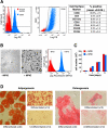
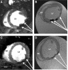
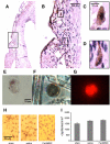
Basic and clinical studies have shown that bone marrow cell therapy can improve cardiac function following infarction. In experimental animals, reported stem cell-mediated changes range from no measurable improvement to the complete restoration of function. In the clinic, however, the average improvement in left ventricular ejection fraction is around 2% to 3%. A possible explanation for the discrepancy between basic and clinical results is that few basic studies have used the magnetic resonance (MR) imaging (MRI) methods that were used in clinical trials for measuring cardiac function. Consequently, we employed cine-MR to determine the effect of bone marrow stromal cells (BMSCs) on cardiac function in rats. Cultured rat BMSCs were characterized using flow cytometry and labeled with iron oxide particles and a fluorescent marker to allow in vivo cell tracking and ex vivo cell identification, respectively. Neither label affected in vitro cell proliferation or differentiation. Rat hearts were infarcted, and BMSCs or control media were injected into the infarct periphery (n = 34) or infused systemically (n = 30). MRI was used to measure cardiac morphology and function and to determine cell distribution for 10 wk after infarction and cell therapy. In vivo MRI, histology, and cell reisolation confirmed successful BMSC delivery and retention within the myocardium throughout the experiment. However, no significant improvement in any measure of cardiac function was observed at any time. We conclude that cultured BMSCs are not the optimal cell population to treat the infarcted heart.
Figures


References
-
- Abbott JD, Huang Y, Liu D, Hickey R, Krause DS, Giordano FJ. Stromal cell-derived factor-1alpha plays a critical role in stem cell recruitment to the heart after myocardial infarction but is not sufficient to induce homing in the absence of injury. Circulation 110: 3300–3305, 2004. - PubMed
-
- Alhadlaq A, Mao JJ. Mesenchymal stem cells: isolation and therapeutics. Stem Cells Dev 13: 436–448, 2004. - PubMed
-
- Amsalem Y, Mardor Y, Feinberg MS, Landa N, Miller L, Daniels D, Ocherashvilli A, Holbova R, Yosef O, Barbash IM, Leor J. Iron-oxide labeling and outcome of transplanted mesenchymal stem cells in the infarcted myocardium. Circulation 116: I38–I45, 2007. - PubMed
-
- Arai T, Kofidis T, Bulte JW, de Bruin J, Venook RD, Berry GJ, McConnell MV, Quertermous T, Robbins RC, Yang PC. Dual in vivo magnetic resonance evaluation of magnetically labeled mouse embryonic stem cells and cardiac function at 1.5 t. Magn Reson Med 55: 203–209, 2006. - PubMed
-
- Assmus B, Honold J, Schachinger V, Britten MB, Fischer-Rasokat U, Lehmann R, Teupe C, Pistorius K, Martin H, Abolmaali ND, Tonn T, Dimmeler S, Zeiher AM. Transcoronary transplantation of progenitor cells after myocardial infarction. N Engl J Med 355: 1222–1232, 2006. - PubMed
Publication types
MeSH terms
Substances
Grants and funding
LinkOut - more resources
Full Text Sources
Other Literature Sources
Medical

