. 2008 Aug 15;112(4):1249-58.
doi: 10.1182/blood-2007-09-114389. Epub 2008 Jun 6.

Antigen-specific T-T interactions regulate CD4 T-cell expansion

Affiliations

Antigen-specific T-T interactions regulate CD4 T-cell expansion

Julie Helft et al. Blood. .

Abstract

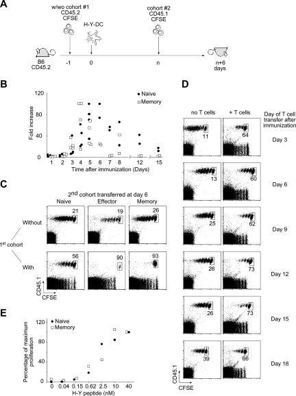
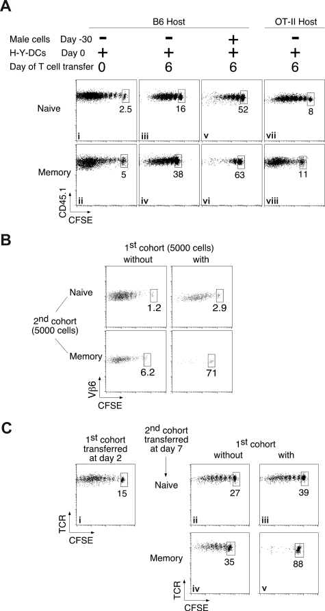
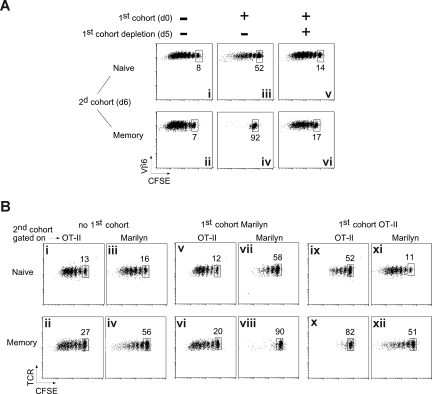
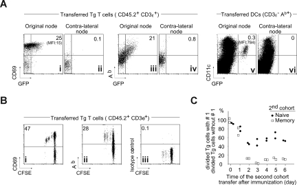
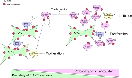
The regulation of CD4 T-cell numbers during an immune response should take account of the amount of antigen (Ag), the initial frequency of Ag-specific T cells, the mix of naive versus experienced cells, and (ideally) the diversity of the repertoire. Here we describe a novel mechanism of T-cell regulation that potentially deals with all of these parameters. We found that CD4 T cells establish a negative feedback loop by capturing their cognate major histocompatibility class (MHC)/peptide complexes from Ag-presenting cells and presenting them to Ag-experienced CD4 T cells, thereby inhibiting their recruitment into the response while allowing recruitment of naive T cells. The inhibition is Ag specific, begins at day 2 (long before Ag disappearance), and cannot be overcome by providing new Ag-loaded dendritic cells. In this way, CD4 T-cell proliferation is regulated in a functional relationship to the amount of Ag, while allowing naive T cells to generate repertoire variety.


Figures

Figure 1
Figure 1
Ag-experienced T-cell proliferation is preferentially inhibited during on ongoing immune response. (A) Two-cohort experimental design. A first cohort of 106 naive CD45.2 CFSE-labeled Marilyn cells was injected intravenously into CD45.2 B6 mice that were immunized by injection of 106 H-Y peptide–loaded LPS-matured DCs into the footpad the next day. After a variable (n) number of days, a second cohort of either 106 naive, 5 × 10⁶ effector, or 2 × 10⁶ memory CD45.1 CFSE-labeled Marilyn cells was injected. CFSE profiles of gated TCR+CD45.1+ cells from the popliteal and inguinal lymph nodes draining the immunization site (DLN) were assessed 6 days after transfer of the second cohort. (B) Kinetics of the response of transferred Marilyn TCR Tg T cells. Naive (106) or memory (2 × 10⁶) CD45.1 CFSE-labeled Marilyn cells were injected intravenously into B6 mice that were primed the next day by injection of 106 H-Y peptide–loaded LPS-matured DCs into the footpad. The percentage of transferred T cells (CD45.1+TCR+) in the draining lymph node was measured at the indicated time points and compared with the percentage measured at day 1 in the absence of antigen. Each dot represents one mouse. Pooled data from 2 independent experiments. (C) Stronger inhibition of Ag-experienced versus naive T-cell proliferation. Naive (left panels), effector (middle panels), or memory (right panels) Marilyn T cells were injected into primed B6 hosts in the absence (top panels) or the presence (bottom panels) of a previous cohort of responding T cells injected 6 days earlier, as in panel A. Dot plots of gated TCR+CD45.1+ cells from the DLN are representative of at least 3 experiments with 2 mice each per group. The percentage of undivided cells, among total Tg Marilyn T cells, is indicated. (D) Antigen persists in vivo at least 18 days. H-Y peptide–loaded LPS-matured DCs were injected into the footpad of female B6 mice, previously injected (right panels) or not (left panels) with naive CD45.2 CFSE-labeled Marilyn LN cells. At the indicated time points, naive CD45.1 CFSE-labeled Marilyn LN cells were transferred intravenously. Dot plots of gated TCR+CD45.1+ cells from the DLN injected at the indicated time after DC priming and analyzed 6 days later. Representative of 2 independent experiments with 2 mice per group for each one. (E) Functional avidity measurement in vivo. Naive (106) or memory (2 × 10⁶) CD45.1 CFSE-labeled Marilyn cells were injected intravenously into B6 mice, which were then primed by injection into the footpad of 106 LPS-matured DCs loaded with the indicated amount of H-Y peptide. Five days later, the number of naive and memory T cells recovered in the DLN was measured.
Figure 2
Figure 2
The preferential inhibition of memory T cells is observed under physiological conditions. (A) Physiologic numbers of responding endogenous polyclonal T cells preferentially inhibit memory T-cell proliferation. Naive (106; top panels) or memory (2 × 10⁶; bottom panels) CD45.1 CFSE-labeled Marilyn cells were injected intravenously into B6 (i-vi) or OT-II (vii,viii) mice immunized by injection of 106 H-Y peptide–loaded LPS-matured DCs into the footpad. (i,ii) Low initial frequencies of endogenous responding T cells do not inhibit naive or memory Marilyn T cells; naive or memory CD45.1 Marilyn T cells were injected on the same day as the H-Y peptide–loaded DCs. (iii-vi) Higher frequencies occurring later in the response inhibit proliferation of newly entering cells. Six days after the initiation of the primary endogenous response by injection of peptide-pulsed DCs into naive B6 hosts (iii,iv), or after a boost of mice that had been immunized 30 days previously (v,vi), new naive or memory CD45.1 CFSE-labeled Marilyn cells were injected, and proliferation was analyzed 6 days later. (vii,viii) Extremely low initial frequencies do not expand to inhibitory numbers in 6 days. Marilyn T cells were injected and analyzed as in panels iii,iv, but into OT-II hosts. Plots are of gated TCR+CD45.1+ cells from the DLN, and are representative of at least 2 experiments with at least 2 mice each per group. (B) Small numbers of responding T cells strongly inhibit small numbers of memory T cells. Five thousand naive (top panels) or memory (bottom panels) CD45.1 Marilyn T cells were injected into primed B6 hosts in the absence (left panels) or the presence (right panels) of a previous cohort of 5000 CD45.2 Marilyn T cells injected 6 days earlier, as in Figure 1A. Pooled draining lymph nodes of 8 to 20 mice were studied. Dot plots of gated TCR+CD45.1+ cells with the percentage of undivided cells are shown. (C) An immune response to endogenous DCs that have processed Ag from a peripheral solid tumor preferentially inhibits memory T-cell proliferation. CD45.2 B6 mice received a transplant of an MCA101 tumor cell line, transfected with the DBY protein. Two days later, they were given a first cohort of CD45.2+ Marilyn T cells (right panels) or not (left panels). Naive (top panels) or memory (bottom panels) CD45.1+ Marilyn T cells were injected 5 days later. Dot plots of gated TCR+CD45.1+ cells from the immunization DLN are representative of at least 3 experiments with 2 mice each per group.
Figure 3
Figure 3
The inhibition is Ag specific and requires the presence of the first cohort of T cells. (A) The inhibition depends on the presence of the first cohort of T cells. Naive (106) or memory (2 × 10⁶) Marilyn T cells were injected into OT-II hosts that had been primed 6 days earlier with H-Y peptide–loaded mature DCs, in the presence (iii-vi) or not (i,ii) of a first cohort of Marilyn-Lat-DTR T cells. The day before the injection of the second cohort, Marilyn-Lat-DTR cells were deleted (v,vi) or not (iii,iv) by the injection of DT. Dot plots of gated CD4+CD45.1+ cells from the DLN studied 6 days later (day 6 + 6) are representative of at least 2 experiments with 2 mice each per group. (B) The inhibition is Ag specific. CD45.1 B6 mice were injected or not (i-iv) with a first cohort of naive CFSE-labeled Marilyn (v-viii) or OT-II (ix-xii) Tg CD4 T cells and were then immunized with LPS-matured DCs pulsed with both OVA and H-Y peptides. Six days later, CFSE-labeled naive (top panels) or memory (bottom panels) OT-II and Marilyn cells were coinjected in the same mouse. Proliferation was measured on the second cohorts (CD45.2+, and Vβ6+ for Marilyn, Vα2hi for OT-II) in the same mouse 6 days later. Representative of 2 independent experiments with 2 mice each per group.
Figure 4
Figure 4
Activated T cells acquire MHC-II molecule during the ongoing immune response in vivo. (A) Acquisition of MHC-II molecules by activated T cells in vivo. Naive Tg CD45.1 Marilyn cells (106) were injected intravenously into B6 mice that were immunized by injection of 106 H-Y peptide–loaded Ab-GFP knockin DCs into the footpad. Two days later, CD69, Ab, and GFP expression were analyzed on the transferred T cells in the draining LN and the nondraining LN (CD45.1+TCR+) by 5-color FACS with doublet exclusion. (B) Proliferating T cells acquire MHC-II in vivo. Naive CFSE-labeled Tg CD45.1 Marilyn cells (106) were injected intravenously into B6 mice and immunized as in panel A. Two days later, CD69 and Ab expression was analyzed in the draining LN on the transferred T cells (CD45.1+TCR+) by 5-color FACS with doublet exclusion. (C) The inhibition starts at day 1 and plateaus by day 2. H-Y peptide–loaded DCs (106) were injected into the footpad of female B6 mice that contained (or not) a first cohort of 106 naive CD45.2 CFSE-labeled Marilyn LN cells. At the indicated time points, a second cohort of 106 CD45.1 CFSE-labeled naive Marilyn LN or 2 × 10⁶ memory Marilyn T cells was injected, and the CFSE profile assessed 6 days later. The percentage of divided TCR+CD45.1+ Marilyn cells in the second cohort is shown for cells in the presence of a first cohort relative to the percentage seen in its absence. Each point is an individual mouse.
Figure 5
Figure 5
Marilyn CD4 T cells capture MHC-II/H-Y in vitro, and induce death of memory but not naive T cells. (A) Purification of MHC-II–bearing T cells. Lymph node CD45.2 Marilyn cells were incubated with H-Y peptide–loaded (100 nM), CFSE-labeled, LPS-matured DCs. Twenty hours later, the T cells were FACS sorted according to CD11c and CFSE expression with exclusion of the doublets. (i) Postsort analysis of the CFSEneg/CD11cneg T-cell fraction. (ii,iii) Expression of Ab by the sorted T cells. (B) T cells that have captured MHC-II/H-Y complexes present them to naive and memory Marilyn T cells. (i,ii) CD45.2 fixed (as described in “In vitro experiments”) naive LN Marilyn cells (0.5 × 10⁶), (iii,iv) 0.5 × 10⁶ purified CD45.2 fixed T cells that had captured MHC-II/H-Y complexes, or (v,vi) 5000 purified H-Y–bearing fresh DCs were used to stimulate 2 × 10⁴ CFSE-labeled CD45.1 naive Marilyn LN cells or negatively purified memory Marilyn T cells. CD69 expression was measured the following day. Representative of 3 experiments. (C) T cells that have captured MHC-II/H-Y complexes suppress memory but not naive Marilyn T-cell proliferation. Purified CD45.2 fixed naive LN Marilyn cells (0.5 × 10⁶), or 0.5 × 10⁶ purified CD45.2 fixed T cells that had captured MHC-II/H-Y complexes (H-Y T cells), or not (unpulsed T cells), and/or various numbers of H-Y–bearing fresh DCs were used to stimulate 2 × 10⁴ CD45.1 naive or memory Marilyn T cells that had been negatively purified by FACS and then CFSE labeled. Proliferation was measured at day 3 by CFSE dilution. Percentage of undivided cells is indicated in each panel, and numbers of recovered cells/well are indicated at the bottom of each panel. Representative of 4 experiments. (D) Fresh DCs, able to stimulate naive cells, cannot relieve inhibition of memory T cells. B6 mice were injected with CD45.2 CFSE-labeled Marilyn cells and were immunized the next day with H-Y peptide–loaded LPS-matured DCs in one hind footpad. Naive or memory CD45.1 CFSE-labeled Marilyn cells were transferred 5 days later. Four hours later, some of the mice (bottom panels) received a second immunization with H-Y peptide–loaded DCs into both the original and the contralateral footpad. The CFSE profile is shown on the second cohort (TCR+ CD45.1+ cells) in the original DLN (left panels) or in the contralateral DLN (right panels) 6 days after they were injected. Representative of 2 experiments with 2 mice per group in each.
Figure 6
Figure 6
Model of CD4 T-cell autoregulation of an immune response. (1) At the onset of the immune response, naive (and also memory in the case of a secondary response, which is not represented here) CD4 T cells are recruited and stimulated by activated APCs displaying antigen. (2) During activation, these CD4 T cells capture the specific MHC-peptide complexes bound by their TCRs. (3) At later time points, the number of activated CD4 T cells that have captured MHC-peptide complexes increases. As these activated Ag-bearing T cells begin to outnumber the APCs, there is increasing probability that newly arriving T cells (and any T cells that have just been activated and need a second hit to continue to divide) will encounter them before encountering a proper professional APC. (4) This T-T interaction leads to inhibition of the Ag-experienced T cells, whereas the normal interaction of naive cells with the professional APCs leads to proliferation.
Figure 7
Figure 7
Ag-presenting Marilyn T cells directly inhibit memory T-cell proliferation in vivo. (A,B) Naive (106) or memory (2 × 10⁶) CD45.1 CFSE-labeled Marilyn T cells were injected intravenously into CD45.2 female hosts. In parallel, CD45.1/2 Marilyn T cells were incubated in vitro overnight with CFSE-labeled H-Y peptide–loaded LPS-matured DCs or with DCs and an anti-TCR antibody. The next day, the 2 sets of in vitro–activated Marilyn T cells were FACS sorted according to CFSE and CD11c expression and injected into the footpad. Six days later, H-Y–loaded DCs were injected into the footpad to stimulate the T cells. CFSE patterns were studied in the DLN 6 days later. Dot plots on gated CD45.1+/CD45.2/Vb6+/CD4+ cells are representative of 2 independent experiments with 2 mice in each.

Similar articles

Cited by

References

    1. Dutton RW, Bradley LM, Swain SL. T cell memory. Annu Rev Immunol. 1998;16:201–223. - PubMed
    1. Ahmed R, Gray D. Immunological memory and protective immunity: understanding their relation. Science. 1996;272:54–60. - PubMed
    1. Sprent J, Surh CD. T cell memory. Annu Rev Immunol. 2002;20:551–579. - PubMed
    1. Merica R, Khoruts A, Pape KA, Reinhardt RL, Jenkins MK. Antigen-experienced CD4 T cells display a reduced capacity for clonal expansion in vivo that is imposed by factors present in the immune host. J Immunol. 2000;164:4551–4557. - PubMed
    1. Badovinac VP, Porter BB, Harty JT. CD8+ T cell contraction is controlled by early inflammation. Nat Immunol. 2004;5:809–817. - PubMed

Publication types

Substances