

Photoreceptor layer topography in children with leber congenital amaurosis caused by RPE65 mutations
- PMID: 18539930
- PMCID: PMC2731624
- DOI: 10.1167/iovs.08-2121
Photoreceptor layer topography in children with leber congenital amaurosis caused by RPE65 mutations
Abstract
Purpose: To study the topography of photoreceptor loss early in the course of Leber congenital amaurosis (LCA) caused by RPE65 mutations.
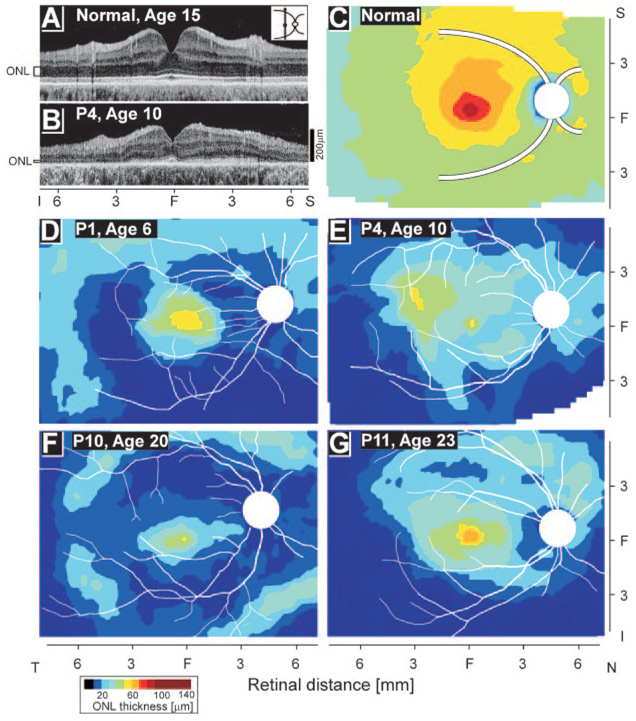
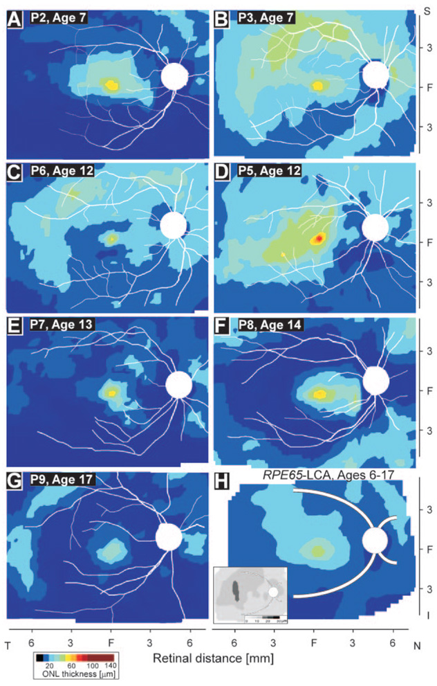
Methods: Young patients with RPE65-LCA (n = 9; ages, 6-17 years) were studied with optical coherence tomography (OCT) in a wide region of central retina. Outer nuclear layer (ONL) thickness was mapped topographically and compared with that in normal subjects and in older patients with RPE65-LCA.
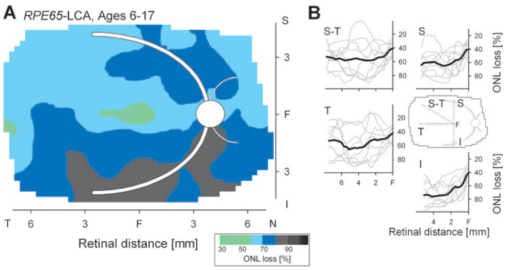
Results: Photoreceptor layer topography was abnormal in all young patients with RPE65-LCA. Foveal and extrafoveal ONL was reduced in most patients. There were interindividual differences, with ONL thicknesses at most retinal locations ranging from near the detectability limit to a significant fraction of normal. These differences were not clearly related to age. In most patients, there was a thinner ONL inferior to the fovea compared with that in the superior retina. Summary maps obtained by aligning and averaging photoreceptor topography across all young patients showed a relative preservation of ONL in the superior-temporal and temporal pericentral retina. These retinal regions also showed the greatest magnitude of interindividual variation.
Conclusions: Photoreceptor loss in the foveal and extrafoveal retina was prominent, even in the youngest patients studied. Differences in the topography of residual photoreceptors in children with RPE65-LCA suggest that it may be advisable to use individualized ONL mapping to guide the location of subretinal injections for gene therapy and thereby maximize the potential for efficacy.
Figures
References
-
- Bainbridge JW, Smith AJ, Barker SS, et al. Effect of gene therapy on visual function in Leber’s congenital amaurosis. N Engl J Med. 2008;358(21):2231–2239. - PubMed
Publication types
MeSH terms
Substances
Grants and funding
LinkOut - more resources
Full Text Sources
Other Literature Sources
Medical
Molecular Biology Databases
Research Materials
