. 2008 Nov;39(5):584-90.
doi: 10.1165/rcmb.2007-0448OC. Epub 2008 Jun 6.

Chronic LPS inhalation causes emphysema-like changes in mouse lung that are associated with apoptosis

Affiliations

Chronic LPS inhalation causes emphysema-like changes in mouse lung that are associated with apoptosis

David M Brass et al. Am J Respir Cell Mol Biol. 2008 Nov.

Abstract

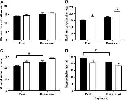
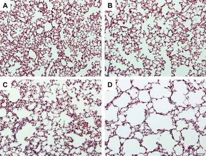
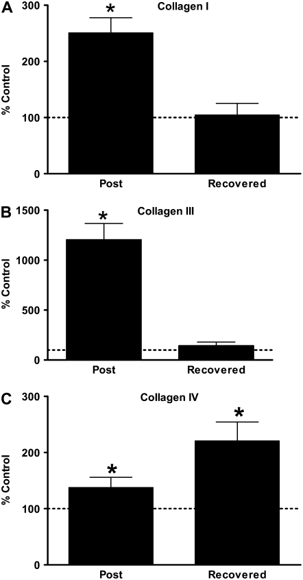
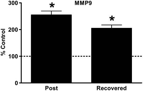
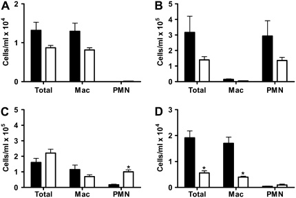
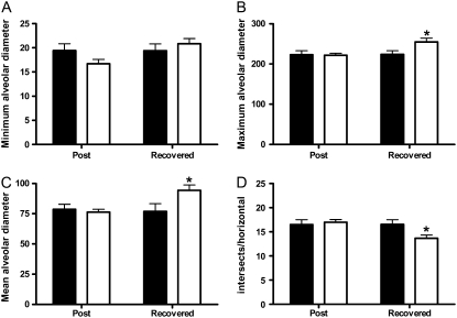
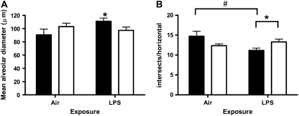
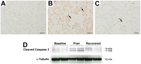
Lipopolysaccharide (LPS) is ubiquitous in the environment. Recent epidemiologic data suggest that occupational exposure to inhaled LPS can contribute to the progression of chronic obstructive pulmonary disease. To address the hypothesis that inhaled LPS can cause emphysema-like changes in mouse pulmonary parenchyma, we exposed C57BL/6 mice to aerosolized LPS daily for 4 weeks. By 3 days after the end of the 4-week exposure, LPS-exposed mice developed enlarged airspaces that persisted in the 4-week recovered mice. These architectural alterations in the lung are associated with enhanced type I, III, and IV procollagen mRNA as well as elevated levels of matrix metalloproteinase (MMP)-9 mRNA, all of which have been previously associated with human emphysema. Interestingly, MMP-9-deficient mice were not protected from the development of LPS-induced emphysema. However, we demonstrate that LPS-induced airspace enlargement was associated with apoptosis within the lung parenchyma, as shown by prominent TUNEL staining and elevated cleaved caspase 3 immunoreactivity. Antineutrophil antiserum-treated mice were partially protected from the lung destruction caused by chronic inhalation of LPS. Taken together, these findings demonstrate that inhaled LPS can cause neutrophil-dependent emphysematous changes in lung architecture that are associated with apoptosis and that these changes may be occurring through mechanisms different than those induced by cigarette smoke.


Figures

<b>Figure 1.</b>
Figure 1.
Chronic lipopolysaccharide (LPS) inhalation challenge causes changes in parenchymal architecture consistent with emphysema in C57BL/6 mouse lung. Quantitative morphometric analysis of C57BL/6 mouse lung histology measuring (A) minimum, (B) maximum, and (C) mean alveolar diameter, and (D) intersects per horizontal. Data are mean ± SEM. *P < 0.05 versus air; #P < 0.05 Post versus Recovered. Solid bars, air; open bars, LPS.
<b>Figure 2.</b>
Figure 2.
Chronic LPS inhalation challenge causes changes in parenchymal architecture consistent with emphysema in C57BL/6 mouse lung. Representative histopathology of C57BL/6 mouse lung exposed to (A and B) filtered air or (C and D) daily LPS inhalation for (A and C) 4 weeks or (B and D) 4 weeks followed by 4 weeks of recovery. Images are ×200 original magnification. Bar = 200 μM.
<b>Figure 3.</b>
Figure 3.
Emphysematous changes in C57BL/6 mouse lung are associated with elevated and persistent procollagen gene expression. Quantitative RT-PCR for (A) pro-α I type I collagen, (B) pro-α III type IV collagen, and (C) pro-α IV type III collagen from mouse whole lung total RNA. Data presented are mean percent of age-matched air control ± SEM. Horizontal line represents 100% of age-matched air control. *P < 0.05 versus age-matched air controls.
<b>Figure 4.</b>
Figure 4.
Emphysematous changes in C57BL/6 mouse lung are associated with elevated and persistent matrix metalloproteinase (MMP)-9 gene expression. Quantitative RT-PCR for MMP-9 from mouse whole lung RNA. Data presented are mean percent of age-matched air control ± SEM. Horizontal line represents 100% of age-matched air control. *P < 0.05 versus age-matched air controls.
<b>Figure 5.</b>
Figure 5.
Acute and chronic LPS inhalation challenge causes substantial neutrophilic inflammation in both C57BL/6 (solid bars) and MMP-9–deficient (open bars) mice. Total cells, macrophages, and neutrophils (PMN) in C57BL/6 and MMP-9–deficient mice (A) at baseline or (B) after a single acute LPS inhalation challenge, or (C) 3 days or (D) 1 month after the end of chronic LPS inhalation challenge. *P < 0.05 versus C57BL/6.
<b>Figure 6.</b>
Figure 6.
Chronic LPS inhalation challenge causes changes in parenchymal architecture independent of MMP-9 in MMP-9–deficient mice. Quantitative morphometric analysis of MMP-9–deficient mouse lung histology measuring (A) minimum, (B) maximum, and (C) mean alveolar diameter, and (D) intersects per horizontal. Data are mean ± SEM. *P < 0.05 versus air. Solid bars, air; open bars, LPS.
<b>Figure 7.</b>
Figure 7.
Emphysematous changes in C3HeB/FeJ mouse lung are partially dependent on the presence of neutrophils. Quantitative morphometric analysis of C3HeB/FeJ mouse lung histology from 1 month recovered animals measuring (A) mean alveolar diameter and (B) intersects per horizontal. Data are mean ± SEM. *P < 0.05 pre-immune serum (solid bars) versus anti-PMN serum (open bars). #P < 0.05 air versus LPS.
<b>Figure 8.</b>
Figure 8.
Emphysematous changes in C57BL/6 mice exposed to chronic LPS inhalation are associated with apoptosis. TUNEL staining in C57BL/6 mouse lung and cleaved caspase 3 Western blot from C57BL/6 mouse lung total protein. (A) Air-exposed at ×100 original magnification; bar = 200 μM. (B) Post at ×400 original magnification; bar = 50 μM. (C) Recovered at ×400 original magnification; bar = 50 μM. (D) Representative cleaved caspase-3 Western blot at baseline and in Post and Recovered groups with α-tubulin as a loading control.

References

    1. Murray CJ, Lopez AD. Alternative projections of mortality and disability by cause 1990–2020: Global burden of disease study. Lancet 1997;349:1498–1504. - PubMed
    1. Khan AJ, Nanchal R. Cotton dust lung diseases. Curr Opin Pulm Med 2007;13:137–141. - PubMed
    1. Haglind P, Rylander R. Exposure to cotton dust in an experimental cardroom. Br J Ind Med 1984;41:340–345. - PMC - PubMed
    1. Castellan RM, Olenchock SA, Kinsley KB, Hankinson JL. Inhaled endotoxin and decreased spirometric values: an exposure-response relation for cotton dust. N Engl J Med 1987;317:605–610. - PubMed
    1. Wang XR, Zhang HX, Sun BX, Dai HL, Hang JQ, Eisen EA, Wegman DH, Olenchock SA, Christiani DC. A 20-year follow-up study on chronic respiratory effects of exposure to cotton dust. Eur Respir J 2005;26:881–886. - PubMed

Publication types