Review
. 2008 Jun 10;26(17):2800-5.
doi: 10.1200/JCO.2007.15.5945.

Liver cancer stem cells

Affiliations
Review

Liver cancer stem cells

Stewart Sell et al. J Clin Oncol. .

Erratum in

  • J Clin Oncol. 2008 Aug 1;26(22): 3819

Abstract

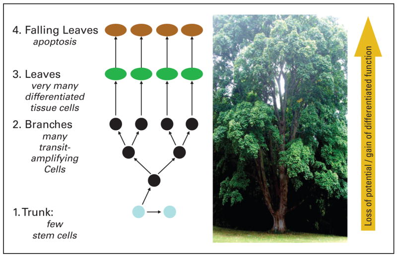
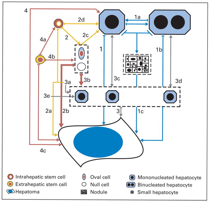
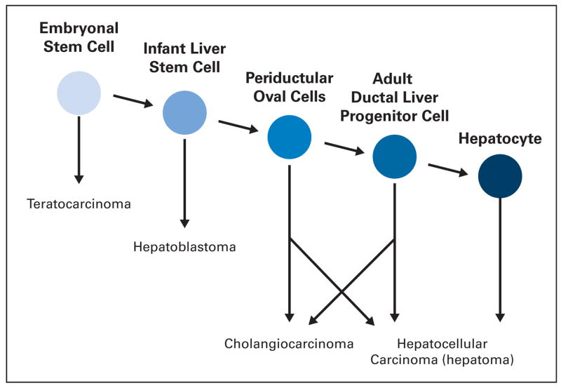
In an effort to review the evidence that liver cancer stem cells exist, two fundamental questions must be addressed. First, do hepatocellular carcinomas (HCC) arise from liver stem cells? Second, do HCCs contain cells that possess properties of cancer stem cells? For many years the finding of preneoplastic nodules in the liver during experimental induction of HCCs by chemicals was interpreted to support the hypothesis that HCC arose by dedifferentiation of mature liver cells. More recently, recognition of the role of small oval cells in the carcinogenic process led to a new hypothesis that HCC arises by maturation arrest of liver stem cells. Analysis of the cells in HCC supports the presence of cells with stem-cell properties (ie, immortality, transplantability, and resistance to therapy). However, definitive markers for these putative cancer stem cells have not yet been found and a liver cancer stem cell has not been isolated.


Figures

Fig 1
Fig 1
Tissue stem cells and normal tissue renewal. The cells in tissue responsible for normal cell turnover are compared with the components of a tree. (1) The stem cells are compared to the base of the tree. They are very few in number, divide rarely, and when they do divide they give rise to one daughter cell that remains a stem cell and one that becomes a transit-amplifying cell. (2) The transit-amplifying cells are like the branches. They divide relatively rapidly as compared with the stem cells. They give rise to additional branches by symmetric division. (3) The differentiated cells of the tissue are like the leaves. They carry out differentiated functions of the tissues. In many tissues the most differentiated cells (eg, cortical neurons [brain]; circulating leukocytes [hematopoietic]; and intestinal epithelium [GI tract]) do not divide. However, in some tissues, particularly the liver, mature hepatocytes are capable of multiple rounds of cell division without losing differentiation function (see text for further discussion). (4) The leaves die by apoptosis (apoptosis = falling out) and are discarded. Deciduous trees discard their dead leaves once a year, whereas human tissues continuously discard dead cells as a part of normal tissue renewal. Modified from Sell.
Fig 2
Fig 2
Carcinogenic lineage model: a role for liver stem or liver progenitor cells. Depicted are four possible pathways from various cells of origin: hepatocytes, intrahepatic stem cells (ductal or periductal), small hepatocytes, or extrahepatic stem cells. Pathway 1, from hepatocytes: hepatomas might develop from mononuclear hepatocytes (pathway 1), binucleated hepatocytes (pathways 1a or 1b), or mature hepatocytes (pathways from preneoplastic nodules derived from hepatocytes are shown as pathway 1c, but are now considered unlikely). Pathway 2, from intrahepatic stem cells either in ducts or next to ducts: directly (pathway 2a) or indirectly (pathway 2b to 2d) from oval or null cell intermediates (pathway 2b) formed from such stem cells. Hepatomas might also develop from large mononucleated hepatocytes derived from oval or null cells (pathway 2c) or directly from intrahepatic stem cells (pathway 2d). Pathway 3, from small hepatocytes: derived from intrahepatic stem cells (pathway 3a), oval or null cells (pathway 3b), mononucleated (pathway 3c) or binucleated hepatocytes (pathway 3d). Pathway 4, from extrahepatic stem cells: directly, or giving rise to other intrahepatic cell types (4a to 4c), or leading to small hepatocytes (pathway 3e). The red arrows indicate likely oval cell to hepatoma lineages (2b and 3b). Modified from Koch and Leffert.
Fig 3
Fig 3
Postulated lineage of liver cells from embryonal cells to mature liver cells and stage of maturation arrest of liver cancers. In the liver, neonatal liver stem cells have more potential than the more differentiated adult liver stem cells and can give rise to hepatoblastomas. In the adult liver, the bipotential liver stem cells in the portal zone (periductal and ductal) can give rise to cholangiocarcinomas, hepatomas, and mixed tumors, whereas mature hepatocytes in the liver lobule can give rise to hepatomas or adenomas. Modified from Sell and Leffert.

Similar articles

Cited by

References

    1. Sell S, Pierce GB. Maturation arrest of stem cell differentiation is a common pathway for the cellular origin of teratocarcinomas and epithelial cancers. Lab Invest. 1994;70:6–22. - PubMed
    1. Sell S. Stem cell origin of cancer and differentiation therapy. Crit Rev Oncology Hematology. 2004;52:1–28. - PubMed
    1. Sell S, Leffert HL. An evaluation of cellular lineages in the pathogenesis of experimental hepatocellular carcinoma. Hepatology. 1982;2:77–86. - PubMed
    1. Sell S, Dunsford H. Evidence for the stem cell origin of hepatocellular carcinoma and cholangiocarcinoma. Am J Pathol. 1989;134:1347–1363. - PMC - PubMed
    1. Sell S. Cellular origin of hepatocellular carcinoma. Cell Develop Biology. 2002;13:419–424. - PubMed

Publication types

MeSH terms