Review
. 2008 Sep 12;283(37):25105-25109.
doi: 10.1074/jbc.R800033200. Epub 2008 Jun 9.

Tethering chemistry and K+ channels

Affiliations
Review

Tethering chemistry and K+ channels

Trevor J Morin et al. J Biol Chem. .

Abstract

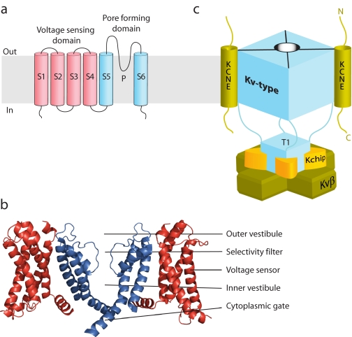
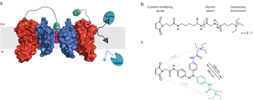
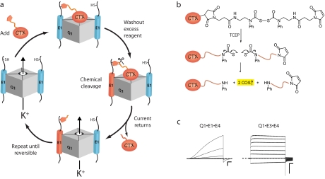
Voltage-gated K+ channels are dynamic macromolecular machines that open and close in response to changes in membrane potential. These multisubunit membrane-embedded proteins are responsible for governing neuronal excitability, maintaining cardiac rhythmicity, and regulating epithelial electrolyte homeostasis. High resolution crystal structures have provided snapshots of K+ channels caught in different states with incriminating molecular detail. Nonetheless, the connection between these static images and the specific trajectories of K+ channel movements is still being resolved by biochemical experimentation. Electrophysiological recordings in the presence of chemical modifying reagents have been a staple in ion channel structure/function studies during both the pre- and post-crystal structure eras. Small molecule tethering agents (chemoselective electrophiles linked to ligands) have proven to be particularly useful tools for defining the architecture and motions of K+ channels. This Minireview examines the synthesis and utilization of chemical tethering agents to probe and manipulate the assembly, structure, function, and molecular movements of voltage-gated K+ channel protein complexes.


Figures

FIGURE 1.
FIGURE 1.
Topological, structural, and schematic model of Kv channel α- and β-subunits. a, topology diagram of a Kv channel. The voltage-sensing domain is shown in red, and the pore-forming domain in blue. b, high resolution structure of the Kv2.1 paddle chimera channel (Protein Data Bank code 2R9R). For clarity, the front and back subunits have been removed. c, schematic model of a Kv-type macromolecular complex. Kv channels that assemble with membrane-embedded KCNE β-subunits do so with a 4:2 stoichiometry. Water-soluble regulatory subunits (Kv channel-interacting proteins (Kchip) and Kvβ) dock onto the N-terminal cytoplasmic tetramerization domain (T1) with a 4:4 stoichiometry. The fourth Kv channel-interacting protein and cytoplasmic C-terminal docking proteins are not visible or shown.
FIGURE 2.
FIGURE 2.
Tethered blockers and inhibitors of K+ channel function. a, Kv channel modified with QA-tethered blockers and a biotin depth charge. The linker on the tethered blocker on the left voltage sensor is long enough to allow the QA to reach its binding site and block conduction, whereas the tethered blocker on the right voltage sensor is too short, and the conductance of the channel is unaffected. Also shown is a membrane-embedded residue that is biotinylated (black triangle) with a linker that permits avidin binding from the extracellular side, but not from the intracellular milieu. The potassium ions are shown in pink, the pore-forming domain in blue, and the voltage-sensing domain in red, and only two Kv subunits are shown for clarity. b, chemical structures of tethered blockers. The effective tether length can be adjusted by incorporating glycine residues Gly(n) using standard solid-phase peptide synthesis: Gly(0), 21 Å; and Gly(7), 45 Å. c, a photoisomerizable azobenzene linker that changes its effective length by ∼7 Å upon exposure to different wavelengths of light.
FIGURE 3.
FIGURE 3.
Tethering strategies with chemically derivatized scorpion toxins (CTX). a, schematic depiction of the iterative counting strategy used to determine the number of E1 subunits in a Q1·K+ channel complex. b, structure and reductive cleavage of CTX-Clv. Tris(2-carboxyethyl)phosphine (TCEP) cleaves the bis(N-phenylcarbamoyl)disulfane linker, giving rise to two secondary amines and 2 eq of carbonyl sulfide gas. c, families of currents from Q1·E1·E4 and Q1·E3·E4 heteromeric K+ channel complexes. Current traces were revealed by subtracting pretreated currents from the currents elicited after washout of CTX-Mal. Command voltages were –100 to 40 mV (20-mV steps). The dashed line indicates zero current. Scale bars = 1 μA and 0.5 s.

References

    1. Bezanilla, F. (2006) Biol. Res. 39 425–435 - PubMed
    1. McCrossan, Z. A., and Abbott, G. W. (2004) Neuropharmacology 47 787–821 - PubMed
    1. Nakajo, K., and Kubo, Y. (2007) J. Gen. Physiol. 130 269–281 - PMC - PubMed
    1. Rocheleau, J. M., and Kobertz, W. R. (2008) J. Gen. Physiol. 131 59–68 - PMC - PubMed
    1. Roepke, T. K., Anantharam, A., Kirchhoff, P., Busque, S. M., Young, J. B., Geibel, J. P., Lerner, D. J., and Abbott, G. W. (2006) J. Biol. Chem. 281 23740–23747 - PubMed

Publication types

LinkOut - more resources