. 2008 Aug;28(16):4975-87.
doi: 10.1128/MCB.00457-08. Epub 2008 Jun 9.

Removal of oxidative DNA damage via FEN1-dependent long-patch base excision repair in human cell mitochondria

Affiliations

Removal of oxidative DNA damage via FEN1-dependent long-patch base excision repair in human cell mitochondria

Pingfang Liu et al. Mol Cell Biol. 2008 Aug.

Abstract

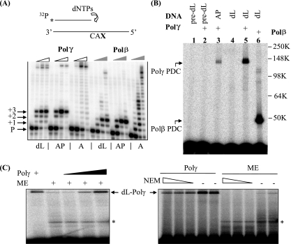
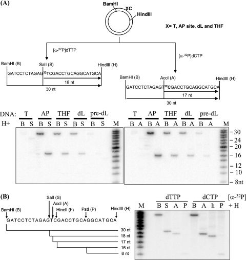
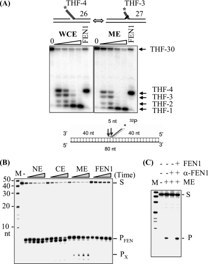
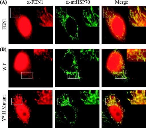
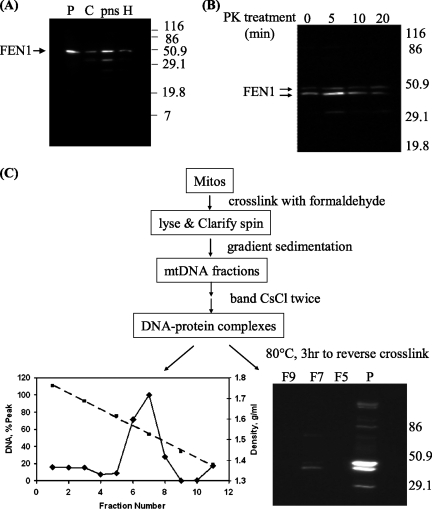
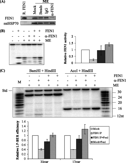
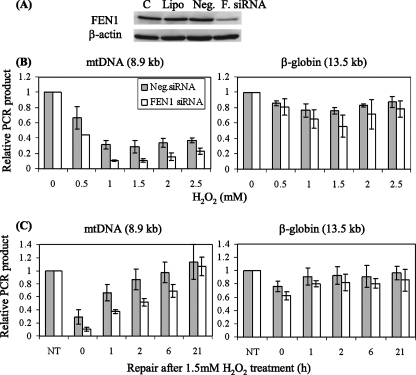
Repair of oxidative DNA damage in mitochondria was thought limited to short-patch base excision repair (SP-BER) replacing a single nucleotide. However, certain oxidative lesions cannot be processed by SP-BER. Here we report that 2-deoxyribonolactone (dL), a major type of oxidized abasic site, inhibits replication by mitochondrial DNA (mtDNA) polymerase gamma and interferes with SP-BER by covalently trapping polymerase gamma during attempted dL excision. However, repair of dL was detected in human mitochondrial extracts, and we show that this repair is via long-patch BER (LP-BER) dependent on flap endonuclease 1 (FEN1), not previously known to be present in mitochondria. FEN1 was retained in protease-treated mitochondria and detected in mitochondrial nucleoids that contain known mitochondrial replication and transcription proteins. Results of immunofluorescence and subcellular fractionation studies were also consistent with the presence of FEN1 in the mitochondria of intact cells. Immunodepletion experiments showed that the LP-BER activity of mitochondrial extracts was strongly diminished in parallel with the removal of FEN1, although some activity remained, suggesting the presence of an additional flap-removing enzyme. Biological evidence for a FEN1 role in repairing mitochondrial oxidative DNA damage was provided by RNA interference experiments, with the extent of damage greater and the recovery slower in FEN1-depleted cells than in control cells. The mitochondrial LP-BER pathway likely plays important roles in repairing dL lesions and other oxidative lesions and perhaps in normal mtDNA replication.


Figures

FIG. 1.
FIG. 1.
The oxidative DNA lesion dL blocks DNA synthesis by Polγ and inactivates the enzyme by forming a covalent DNA-protein cross-link during attempted SP-BER. (A) A running-start experiment was performed to investigate the effect of dL or an AP site on Polγ-mediated DNA synthesis. The reactions were performed with proofreading-deficient Polγ holoenzyme (indicated by open wedges) and Polβ (indicated by filled wedges). In the schematic drawing, CAX in the template strand indicates the sequence: C and A nucleotides followed by an abasic residue (dL or AP) or an undamaged adenylate nucleotide (A). P, +1, +2, and +3 indicate, respectively, the labeled primer and the extension products with one, two, or three additional nucleotides. The full-length extension products appear near the top of the images and are most readily seen with the undamaged A substrate (see designations below the panel). dNTPs, dinucleotide triphosphates. (B) dL traps purified Polγ. A 3′-end-labeled, nicked-DNA duplex with a 5′ dL in the gap was used to mimic the repair intermediate after Ape1 incision. The protein-DNA complex (PDC) of Polγ formed with an incised AP site and reductive trapping with NaBH4 (37) is shown in lane 3. pre-dL, the dL precursor. Molecular weights in thousands (K) are listed on the right of the gel. (C) dL traps Polγ in ME. A protein-DNA cross-link band was detected with ME of HeLa cells and migrated identically to the recombinant Polγ marker, which was also titrated into the extract for the right three lanes of the left panel. Upon treatment with N-ethylmaleimide (NEM; 5, 10, and 20 mM), this high-molecular-weight band disappeared, consistent with the sensitivity of Polγ to this reagent. The identity of the lower band seen with ME (indicated by an asterisk) is unknown, and it may result from dL cross-linking with contaminating Polβ, a proteolytic fragment of Polγ, or other unidentified mitochondrial proteins with dRP lyase activity.
FIG. 2.
FIG. 2.
Repair of dL in ME via LP-BER. (A) Plasmid DNA containing a site-specific unmodified base or abasic lesion (X) was constructed and used for repair as described previously (51) (designated by X followed by an unmodified C nucleotide). (Left panel) Repair (via both SP-BER and LP-BER) was monitored by the incorporation of [α-32P]dTMP into the plasmid, and DNA fragments were revealed after digestion with different restriction endonuclease sets (BamHI plus HindIII and SalI plus HindIII). (Right panel) LP-BER was monitored exclusively by the incorporation of [α-32P]dCMP, and the repaired DNA fragments were revealed by treatment with restriction endonuclease sets of BamHI plus HindIII and AccI plus HindIII. In both panels, lanes showing incubations with the dL precursor (pre-dL) confirm a lack of repair in the absence of a BER substrate. (B) LP-BER was confirmed by treatment with different combinations of restriction enzymes. Strand displacement was ruled out as the mechanism of radioactive nucleotide incorporation because, in such a case, an equal incorporation would be expected for every C position downstream of the abasic residue.
FIG. 3.
FIG. 3.
Flap endonuclease activity in human ME. (A) Flap endonuclease activities in GM1310 whole-cell extracts (WCE) and ME. A flap-containing DNA duplex was used to mimic a repair intermediate of LP-BER. The 4-nt flap (with a 5′-terminal THF residue) substrate equilibrates between two structures: a 5′ THF-4-nt single flap and a double flap of 1 nt on the 3′ side and THF-3 nt on the 5′ side. The numbers along the right side indicate the product oligonucleotide sizes (nt). Note that the presence of a 5′ THF residue at the flap ends contributes mobility of <1-nt equivalent. (B) Flap endonuclease activity in HeLa ME. NE, CE, or ME (100 ng each) or 0.1 ng purified FEN1 was incubated with 1 pmol flap substrate at 30°C for 5, 10, 20, 40, and 60 min as indicated by the wedges; the second lane (left panel) corresponds to incubation of the substrate for 60 min without added protein. S indicates the uncleaved substrate, and PFEN and PX indicate the products of FEN1 and nonspecific exonuclease, respectively. M, molecular size marker. (C) FEN1 was depleted from ME with agarose beads conjugated with antibody against FEN1 (α-FEN1). Flap endonuclease activity was assayed with 100 ng ME, FEN1-depeleted ME, or FEN1-depleted ME supplemented with 0.1 ng purified FEN1. M, molecular size markers; S, uncleaved substrate; P, cleavage product of FEN1.
FIG. 4.
FIG. 4.
Immunolocalization of FEN1 to mitochondria. (A) Representative images of colocalization of FEN1 and mitochondrion-specific heat shock protein (mtHSP70) in HeLa cells. Fixed HeLa cells were costained with anti-FEN1 (α-FEN1) and anti-mtHsp70 (α-mtHSP70) antibodies. In the merged views, the yellow spots indicate colocalization of FEN1 and mtHsp70. The boxed area in each frame is shown magnified in the upper right; this better illustrates the extranuclear staining of FEN1 and mtHSP70. (B) Representative images of the localization of FEN1 and mtHsp70 in HeLa cells expressing wild-type (WT) and Y83H mutant forms of the nuclease.
FIG. 5.
FIG. 5.
FEN1 in protease-treated mitochondria and in nucleoids. Molecular weights (in thousands) are given to the side of the gels. (A) FEN1 was detected in purified mitochondria. H, cell homogenate; pns, postnuclear supernatant; C, cytosol fraction; P: purified mitochondria. (B) FEN1 in mitochondria is resistant to proteinase K (PK) treatment. Pure mitochondria were treated with proteinase K for 0, 5, 10, and 20 min before lysis, and 5 μg of mitochondrial lysate was loaded onto each lane for WB. (C) FEN1 is localized in mitochondrial DNA nucleoids. Formaldehyde cross-linked mitochondrial DNA nucleoids were purified as shown in the flow chart. Mitos, mitochondria. After cross-links were reversed, DNA and FEN1 protein in each CsCl fraction were detected by fluorescence and immunoblotting, respectively. F5, F7, and F9, CsCl fractions 5, 7, and 9, respectively; P, purified mitochondria.
FIG. 6.
FIG. 6.
LP-BER in mitochondria dependent on FEN1. α-FEN1, FEN1 antibody. (A) Western blot showing immunodepletion of FEN1 from GM1310 ME, with mtHsp70 as a loading control. ME (20 μg) was loaded onto each lane. R. FEN1, purified recombinant FEN1; IgG, immunoglobulin G. (B) Diminished flap endonuclease activity following FEN1 immunodepletion. The substrate used was the THF-4-nt flap (see Fig. 3A). The strong band at the bottom of lane 2 is likely due to the 5′-3′ exonuclease activity of FEN1. (C) LP-BER assay with a THF substrate was performed in the presence of [α-32P]dCTP at 30°C for 30 min. Repair products were revealed after digestion with restriction endonuclease sets BamHI plus HindIII and AccI plus HindIII, which released a 30-nt fragment and a 17-nt fragment, respectively, from the plasmid DNA. The relative LP-BER efficiency of each sample was calculated by normalizing the radioactivity of the 30-nt or the 17-nt fragment to that of the 35-nt internal standard (Std). The internal standard was added after reactions were terminated to control for material loss during phenol extraction and ethanol precipitation. For reactions supplemented with exogenous FEN1, 20 ng of purified FEN1 was added to 12 μg of ME. The experiments were repeated three times. The average values and standard deviations are shown in a bar diagram. M, molecular size marker. IP, immunoprecipitation.
FIG. 7.
FIG. 7.
Suppression of FEN1 interferes with efficient repair of oxidative damage in mtDNA. (A) FEN1 was knocked down in HeLa cells by using siRNA. C, controls; Lipo, cells treated with Lipofectamine 2000; Neg., negative control siRNA; F. siRNA, FEN1-specific siRNA. (B and C) DNA damage and repair were assayed by long-range QPCR generating an 8.9-kb product from mtDNA and a 13.5-kb product from the β-globin gene for nuclear DNA. These were normalized to the total DNA in the sample for the nuclear product or a 221-bp PCR product for the mtDNA signal. (B) Damage was induced by various doses of H2O2 for 30 min. (C) Repair of mtDNA and nuclear DNA was monitored by QPCR after 30 min of 1.5 mM H2O2 treatment. NT, cells not treated with H2O2. Results shown are averages of three independent siRNA transfections; error bars represent standard deviations.

References

    1. Akbari, M., T. Visnes, H. E. Krokan, and M. Otterlei. 2008. Mitochondrial base excision repair of uracil and AP sites takes place by single-nucleotide insertion and long-patch DNA synthesis. DNA Repair (Amsterdam) 7605-616. - PubMed
    1. Bender, A., K. J. Krishnan, C. M. Morris, G. A. Taylor, A. K. Reeve, R. H. Perry, E. Jaros, J. S. Hersheson, J. Betts, T. Klopstock, R. W. Taylor, and D. M. Turnbull. 2006. High levels of mitochondrial DNA deletions in substantia nigra neurons in aging and Parkinson disease. Nat. Genet. 38515-517. - PubMed
    1. Bogenhagen, D. F. 1999. Repair of mtDNA in vertebrates. Am. J. Hum. Genet. 641276-1281. - PMC - PubMed
    1. Bogenhagen, D. F., D. Rousseau, and S. Burke. 2008. The layered structure of human mitochondrial DNA nucleoids. J. Biol. Chem. 2833665-3675. - PubMed
    1. Bohr, V. A. 2002. Repair of oxidative DNA damage in nuclear and mitochondrial DNA, and some changes with aging in mammalian cells. Free Radic. Biol. Med. 32804-812. - PubMed

Publication types