. 2008 Jun 16;181(6):893-901.
doi: 10.1083/jcb.200712028. Epub 2008 Jun 9.

Mps1 kinase activity restrains anaphase during an unperturbed mitosis and targets Mad2 to kinetochores

Affiliations

Mps1 kinase activity restrains anaphase during an unperturbed mitosis and targets Mad2 to kinetochores

Anthony Tighe et al. J Cell Biol. .

Abstract

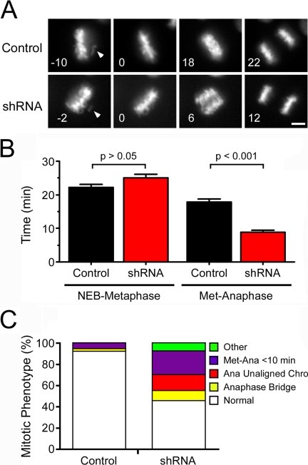
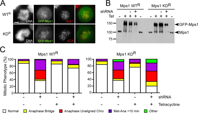
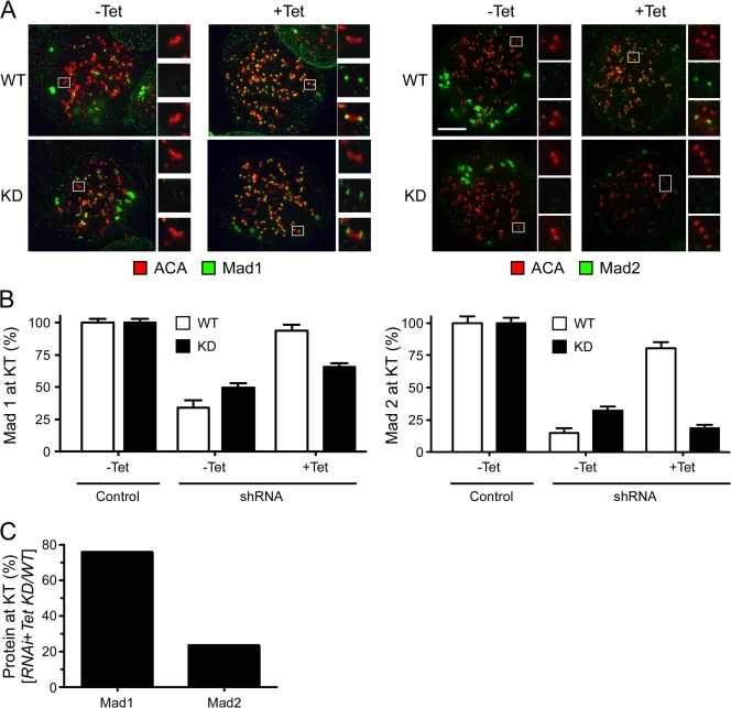
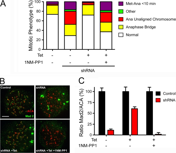
Mps1 is an upstream component of the spindle assembly checkpoint, which, in human cells, is required for checkpoint activation in response to spindle damage but not apparently during an unperturbed mitosis. Mps1 also recruits Mad1 and Mad2 to kinetochores. However, whether the enzymatic activity of Mps1 is required for these processes is unclear. To address this question, we established an RNA interference (RNAi) complementation assay. Repression of Mps1 triggers premature anaphase, often with unaligned or maloriented chromosomes. This phenotype is rescued by an RNAi-resistant wild-type Mps1 transgene but not by a catalytically inactive mutant. An analogue-sensitive allele, Mps1(M602A), also rescues the RNAi-induced defect, but not when inhibited by the adenosine triphosphate analogue 1-NM-PP1. Thus, Mps1 activity does restrain anaphase during an unperturbed mitosis. Furthermore, although catalytically inactive Mps1 can restore kinetochore localization of Mad1, only the active kinase restores Mad2 localization. Thus, in human cells, Mps1 catalytic activity is required for spindle checkpoint function and recruitment of Mad2.


Figures

Figure 1.
Figure 1.
Mps1 is required for spindle checkpoint function during an unperturbed mitosis. HeLa cells cotransfected with control or Mps1 shRNA plus a GFP–histone H2B construct were analyzed by time-lapse microscopy. (A) Image sequences showing the time in minutes from when the last chromosome aligns (arrowheads) to anaphase onset. (B) Bar graph quantifying the time taken from NEB to metaphase and from metaphase to anaphase in control and Mps1-deficient cells. Values represent the mean and SEM (error bars) derived from at least 62 cells. (C) Bar graph quantifying mitotic abnormalities observed in control or Mps1-deficient cells. Bar, 5 μm.
Figure 2.
Figure 2.
Mps1 kinase activity is required for spindle checkpoint function. HeLa cells expressing tet-inducible RNAi-resistant GFP-tagged Mps1 fusions were transfected with shRNA vectors and analyzed by immunofluorescence microscopy, immunoblotting, and time-lapse microscopy. (A) Immunofluorescence images showing kinetochore localization of wild-type Mps1 (WTR) and the catalytically inactive D664A mutant (KDR). (B) Immunoblot showing repression of the endogenous Mps1 and induction of the RNAi-resistant GFP-tagged proteins. (C) Bar graph quantifying the mitotic phenotypes observed after repression of endogenous Mps1; whereas Mps1 WTR rescues the RNAi defect, Mps1 KDR exacerbates the phenotype. At least 132 cells were scored in each category. Bar, 5 μm.
Figure 3.
Figure 3.
Mps1 kinase activity is required for kinetochore localization of Mad2. HeLa cells expressing tet-inducible RNAi-resistant GFP-tagged Mps1 fusions were transfected with shRNA vectors, treated with nocodazole for 2 h, and analyzed to detect centromeres (ACA; red) and Mad1 or Mad2 (green). (A) Deconvolved image stacks showing that although Mps1 WTR restores Mad2 localization, Mps1 KDR does not. Enlargements of highlighted (boxed) kinetochores show the presence or absence of either Mad1 or Mad2. (B) Bar graph quantifying Mad1 and Mad2 pixel intensities at kinetochores normalized to the ACA signal. Values represent the mean and SEM (error bars) derived from at least 106 kinetochores from at least nine cells. (C) Bar graph plotting the amount of Mad1 and Mad2 at kinetochores in cells reconstituted with Mps1 KDR as a percentage of those reconstituted with Mps1 WTR. Bar, 5 μm.
Figure 4.
Figure 4.
Chemical genetic inhibition of Mps1 kinase activity inhibits the checkpoint and Mad2 localization. HeLa cells expressing tet-inducible RNAi-resistant GFP-tagged Mps1 fusions were transfected with shRNA vectors and analyzed by time-lapse microscopy and immunofluorescence. (A) Bar graph quantifying the mitotic phenotypes observed after repression of endogenous Mps1; the ability of Mps1M602A to rescue the RNAi defect is reversed by 1-NM-PP1. (B) Projections of deconvolved image stacks of cells stained to detect centromeres (ACA; red) and Mad2 (green) showing that the ability of Mps1M602A to restore kinetochore localization of Mad2 is reversed by 1-NM-PP1. (C) Bar graph quantifying Mad2 pixel intensities at kinetochores normalized to the ACA signal. Values represent the mean and SEM (error bars) derived from at least 64 kinetochores from at least four cells. Bar, 5 μm.

References

    1. Abrieu, A., L. Magnaghi-Jaulin, J.A. Kahana, M. Peter, A. Castro, S. Vigneron, T. Lorca, D.W. Cleveland, and J.C. Labbe. 2001. Mps1 is a kinetochore-associated kinase essential for the vertebrate mitotic checkpoint. Cell. 106:83–93. - PubMed
    1. Baumann, C., R. Korner, K. Hofmann, and E.A. Nigg. 2007. PICH, a centromere-associated SNF2 family ATPase, is regulated by Plk1 and required for the spindle checkpoint. Cell. 128:101–114. - PubMed
    1. Campbell, M.S., G.K. Chan, and T.J. Yen. 2001. Mitotic checkpoint proteins HsMAD1 and HsMAD2 are associated with nuclear pore complexes in interphase. J. Cell Sci. 114:953–963. - PubMed
    1. Clute, P., and J. Pines. 1999. Temporal and spatial control of cyclin B1 destruction in metaphase. Nat. Cell Biol. 1:82–87. - PubMed
    1. Dorer, R.K., S. Zhong, J.A. Tallarico, W.H. Wong, T.J. Mitchison, and A.W. Murray. 2005. A small-molecule inhibitor of Mps1 blocks the spindle-checkpoint response to a lack of tension on mitotic chromosomes. Curr. Biol. 15:1070–1076. - PubMed

Publication types

MeSH terms