. 2008 Jun 16;181(6):885-92.
doi: 10.1083/jcb.200803042. Epub 2008 Jun 9.

Centromere mitotic recombination in mammalian cells

Affiliations

Centromere mitotic recombination in mammalian cells

Isabel Jaco et al. J Cell Biol. .

Abstract

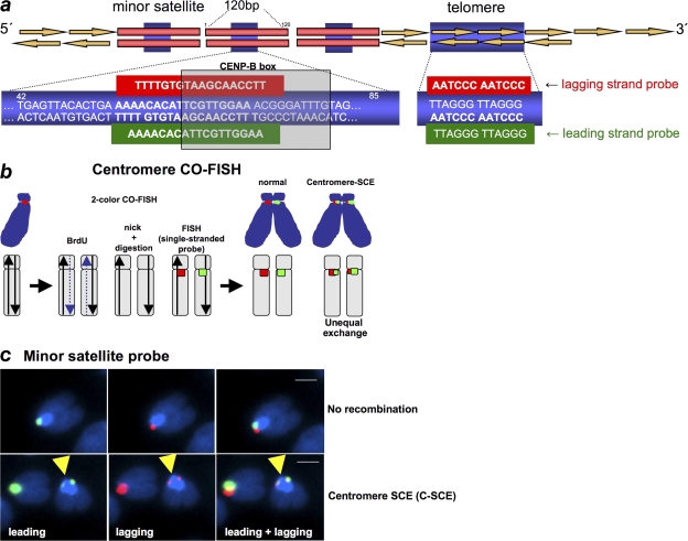
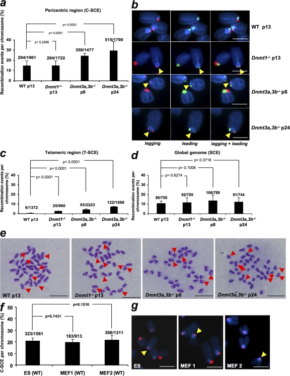
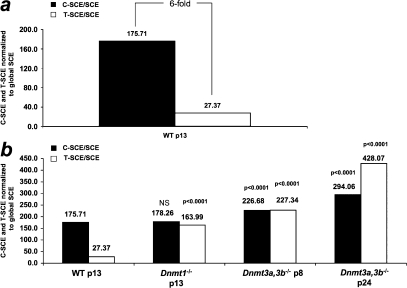
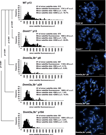
Centromeres are special structures of eukaryotic chromosomes that hold sister chromatid together and ensure proper chromosome segregation during cell division. Centromeres consist of repeated sequences, which have hindered the study of centromere mitotic recombination and its consequences for centromeric function. We use a chromosome orientation fluorescence in situ hybridization technique to visualize and quantify recombination events at mouse centromeres. We show that centromere mitotic recombination occurs in normal cells to a higher frequency than telomere recombination and to a much higher frequency than chromosome-arm recombination. Furthermore, we show that centromere mitotic recombination is increased in cells lacking the Dnmt3a and Dnmt3b DNA methyltransferases, suggesting that the epigenetic state of centromeric heterochromatin controls recombination events at these regions. Increased centromere recombination in Dnmt3a,3b-deficient cells is accompanied by changes in the length of centromere repeats, suggesting that prevention of illicit centromere recombination is important to maintain centromere integrity in the mouse.


Figures

Figure 1.
Figure 1.
CO-FISH to measure recombination rates at minor satellite sequences. (a) A scheme of mouse minor satellite repeats and telomere repeats is shown indicating the position of the CENP-B box (bp 62–78). PNA probes are also indicated. (b) A diagram explaining the CO-FISH procedure is shown. Cells are allowed to replicate once in the presence of BrdU, giving rise to chromosomes with one BrdU-containing chromatid (dashed line). The BrdU-containing DNA strand is digested and single-stranded minor satellite probes against the lagging (red) or the leading (green) strand are hybridized to the remaining non–BrdU-labeled strand. Cen-CO-FISH labels centromeric minor satellite sequences from the lagging or leading strand. An SCE within minor satellite sequences (C-SCE) will lead to two unequal signals per chromosome after hybridization with either the leading or lagging single-stranded probes. (c) Representative Cen-CO-FISH images showing no recombination (top) or a C-SCE after hybridization with the leading and lagging minor satellite PNA probes (bottom). Bars, 1 μm.
Figure 2.
Figure 2.
Recombination events per chromosome at the indicated chromosomal regions in wild-type and Dnmt-deficient ES cells. (a) Error bars correspond to two independent experiments (n = 2). The total number of C-SCE detected out of the total number of chromosomes analyzed per genotype is also indicated on top of each bar. (b) Representative examples of C-SCE in ES cells of the indicated passage and genotype. Yellow arrows point to the C-SCE event. Bars, 3 μm. (c) T-SCE data were obtained from Gonzalo et al. (2006). (d) Quantification of global SCE events in wild-type and Dnmt-deficient mouse ES cells at the indicated passage number. One culture of each genotype was used for the analysis. The total number of SCE events out of the total number of chromosomes analyzed is indicated on top of each bar. (e) Representative images of SCE events. The arrows indicate chromosomes showing SCE events. Bars, 10 μm. (f) Quantification of C-SCE frequencies in mouse wild-type ES cells and two independent wild-type MEF cells. No significant differences were observed between the indicated cells. Numbers above bars represent total C-SCE events out of the total number of chromosomes counted. (g) Representative CO-FISH images after labeling lagging (red) strand centromeres are shown. Yellow arrows indicate C-SCE events. Bars, 5 μm. Error bars represent standard error.
Figure 3.
Figure 3.
Centromeres are more recombinogenic than telomeres. (a and b) Direct comparison of recombination events at centromeres and telomeres per kilobase relative to global SCE events in kilobase (Tables I–III) in wild-type and Dnmt-deficient ES cells. Note that centromeres are sixfold more recombinogenic than telomeres. The T-SCE/SCE and C-SCE/SCE ratios also indicate that both telomeres and centromeres are more recombinogenic than the global genome. C-SCE and T-SCE, but not SCE (see Fig. 2d), are further increased in the absence of Dnmts. Statistical significance comparisons with the respective wild-type value are indicated on top of each bar.
Figure 4.
Figure 4.
Quantification of minor satellite length in wild-type and Dnmt-deficient ES cells. Minor satellite fluorescence distribution in arbitrary units of fluorescence (a.u.f) of wild-type (wt), Dnmt1−/−, and Dnmt3a,3b−/− ES cells at the indicated passage number as determined by Q-FISH using a mouse minor satellite PNA probe (left). Representative images of metaphase spreads of the indicated genotypes are also shown (right). A nonparametric ANOVA test, the Kruskall-wallis test (P < 0.0001), indicated that the observed differences in the centromere fluorescence were not because of coincidences or random sampling. Furthermore, a Dunn's post test indicated that the differences were significant (P < 0.001) for each pair of comparisons. The p-values in the figure refer to comparisons of the different genotypes with wild-type cells. Bars, 7 μm.

Similar articles

Cited by

References

    1. Bailey, S.M., E.H. Goodwin, J. Meyne, and M.N. Cornforth. 1996. CO-FISH reveals inversions associated with isochromosome formation. Mutagenesis. 11:139–144. - PubMed
    1. Bailey, S.M., M.A. Brenneman, and E.H. Goodwin. 2004. Frequent recombination in telomeric DNA may extend the proliferative life of telomerase-negative cells. Nucleic Acids Res. 32:3743–3751. - PMC - PubMed
    1. Bechter, O.E., J.W. Shay, and W.E. Wright. 2004. The frequency of homologous recombination in human ALT cells. Cell Cycle. 3:547–549. - PubMed
    1. Benetti, R., S. Gonzalo, I. Jaco, G. Schotta, P. Klatt, T. Jenuwein, and M.A. Blasco. 2007. Suv4-20h deficiency results in telomere elongation and derepression of telomere recombination. J. Cell Biol. 178:925–936. - PMC - PubMed
    1. Blackburn, E.H. 2001. Switching and signalling at the telomere. Cell. 106:661–673. - PubMed

Publication types

Substances