. 2008 Aug;52(2):256-63.
doi: 10.1161/HYPERTENSIONAHA.108.112706. Epub 2008 Jun 9.

Role of inflammation in the development of renal damage and dysfunction in angiotensin II-induced hypertension

Affiliations

Role of inflammation in the development of renal damage and dysfunction in angiotensin II-induced hypertension

Tang-Dong Liao et al. Hypertension. 2008 Aug.

Abstract

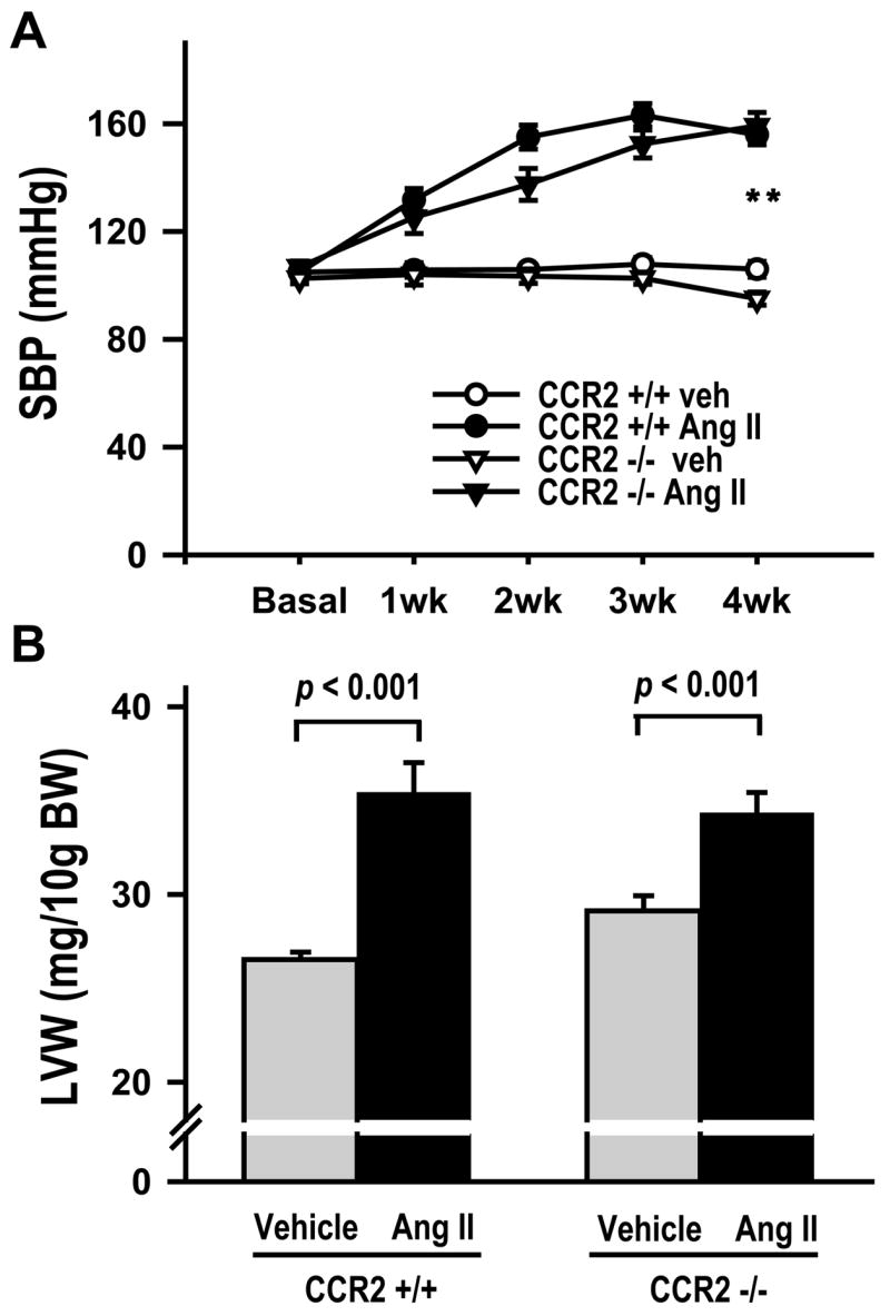
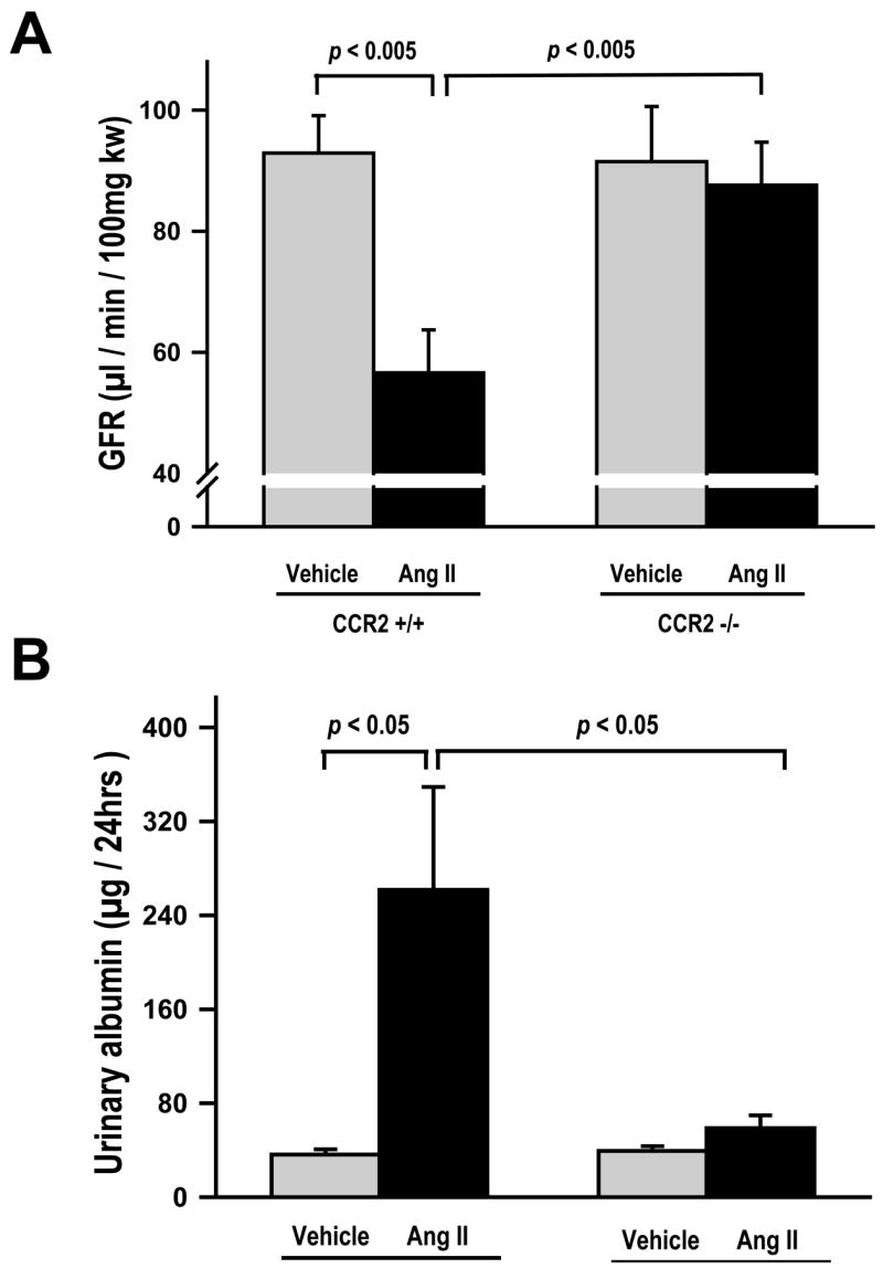
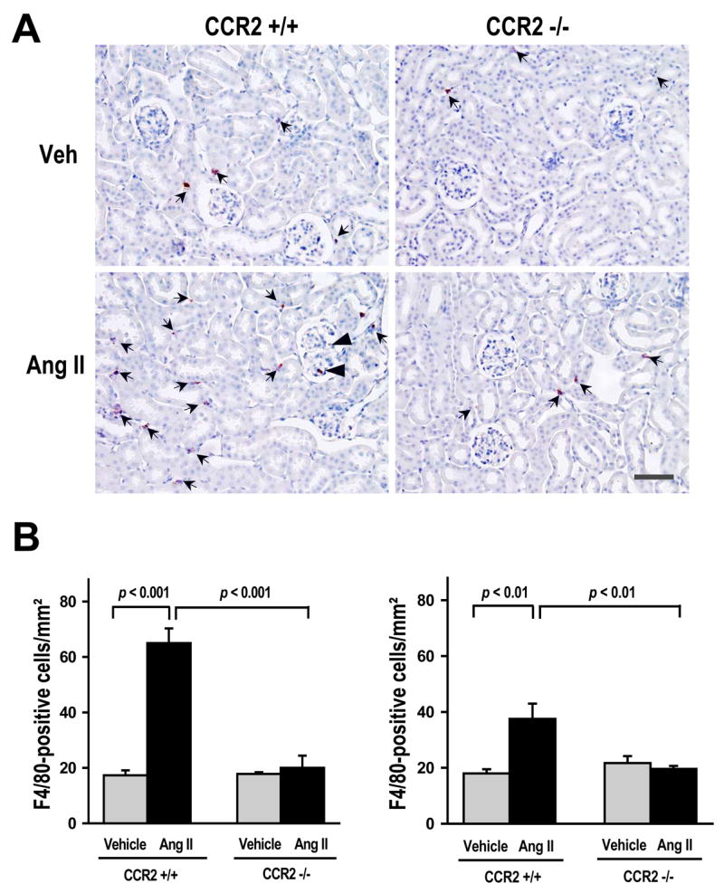
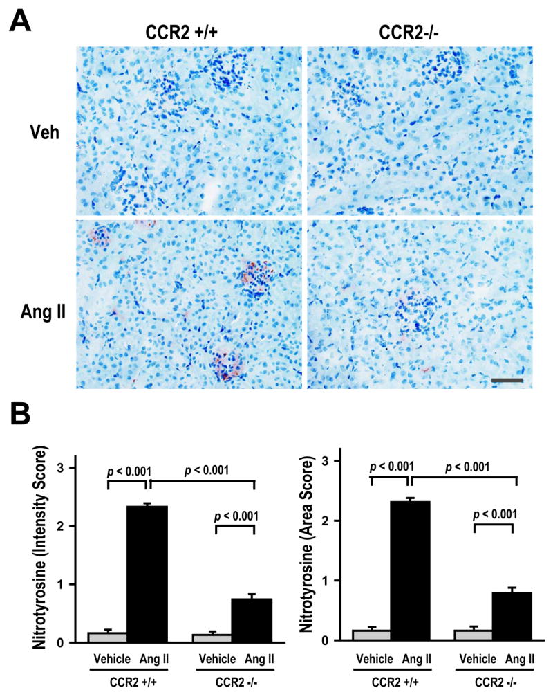
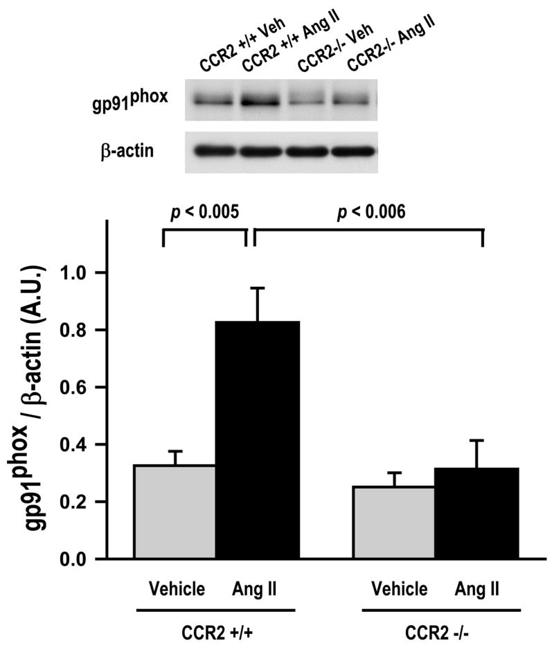
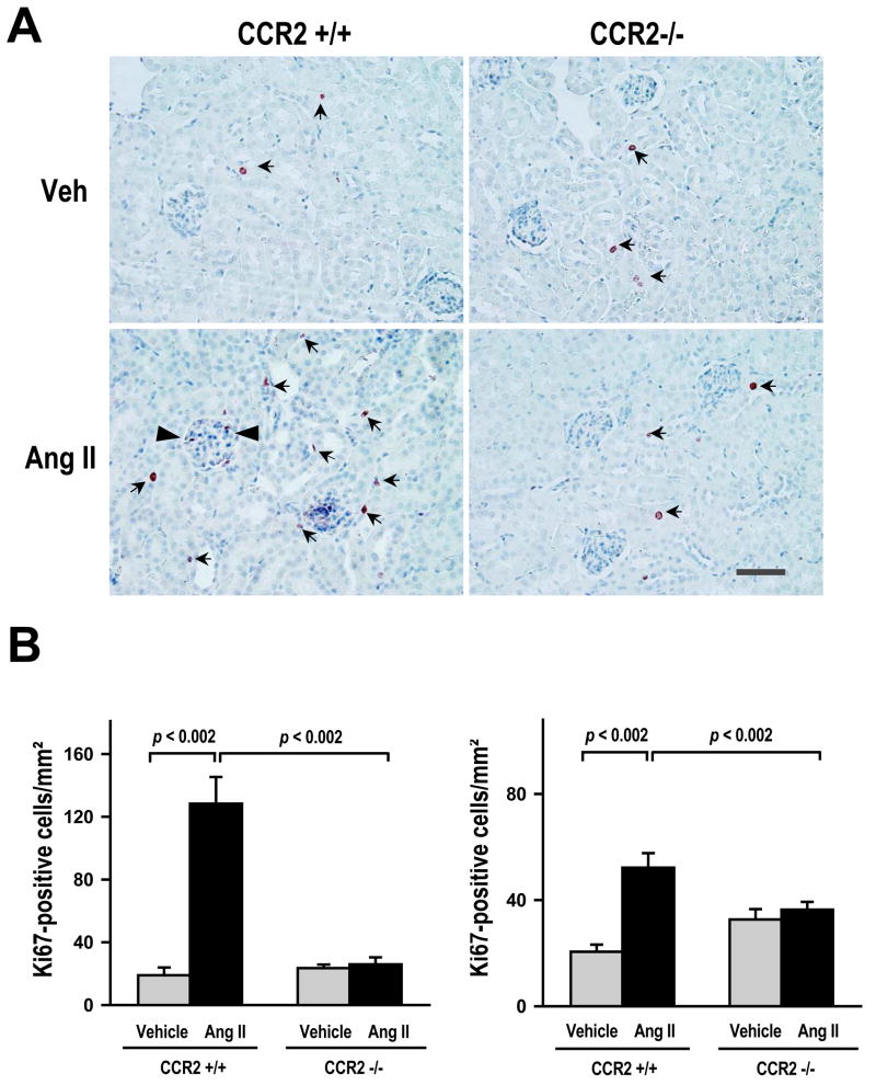
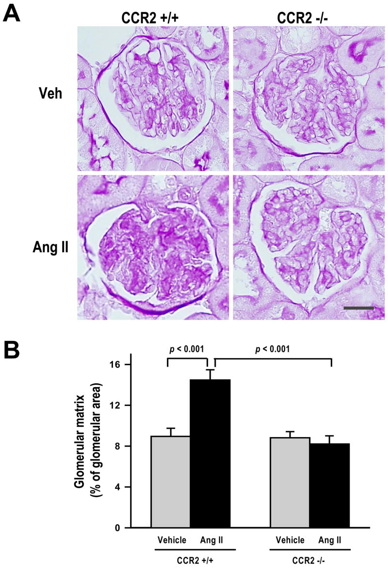
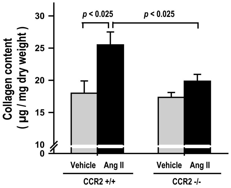
Angiotensin II (Ang II)-induced hypertension is associated with an inflammatory response that may contribute to the development of target organ damage. We tested the hypothesis that, in Ang II-induced hypertension, CC chemokine receptor 2 (CCR2) activation plays an important role in the development of renal fibrosis, damage, and dysfunction by causing oxidative stress, macrophage infiltration, and cell proliferation. To test this hypothesis, we used CCR2 knockout mice (CCR2-/-). The natural ligand of CCR2 is monocyte chemoattractant protein-1, a chemokine important for macrophage recruitment and activation. CCR2-/- and age-matched wild-type (CCR2+/+) C57BL/6J mice were infused continuously with either Ang II (5.2 ng/10 g per minute) or vehicle via osmotic minipumps for 2 or 4 weeks. Ang II infusion caused similar increases in systolic blood pressure and left ventricular hypertrophy in both strains of mice. However, in CCR2-/- mice with Ang II-induced hypertension, oxidative stress, macrophage infiltration, albuminuria, and renal damage were significantly decreased, and glomerular filtration rate was significantly higher than in CCR2+/+ mice. We concluded that, in Ang II-induced hypertension, CCR2 activation plays an important role in the development of hypertensive nephropathy via increased oxidative stress and inflammation.


Figures

Figure 1
Figure 1
(A) Systolic blood pressure (SBP), and (B) Left ventricular weight (LVW) corrected for body weight (BW) in CCR2+/+ and CCR2−/− mice infused with either vehicle or Ang II. SBP was similar in vehicle groups of both strains. Ang II infusion increased SBP and LVW significantly but similarly in both strains. ** p < 0.001, CCR2+/+ and CCR2−/− vehicle vs Ang II; n = 11 to 13 per group.
Figure 2
Figure 2
(A) Glomerular filtration rate (GFR), and (B) urinary albumin in CCR2+/+ and CCR2−/− mice infused with either vehicle or Ang II. GFR was similar in vehicle groups of both strains. In CCR2+/+ with Ang II-induced hypertension, GFR was significantly lower compared to vehicle (p<0.005, vehicle vs Ang II) and also compared to CCR2−/− with Ang II-induced hypertension (p < 0.005, CCR2+/+ vs CCR2−/−). In CCR2−/− with Ang II-induced hypertension, GFR did not change (NS; vehicle vs Ang II), (n = 11 to 13 per group). Urinary albumin excretion increased significantly in CCR2+/+ with Ang II-induced hypertension (p < 0.05, vehicle vs Ang II), while in CCR2−/−, it did not change. (n = 8 to 12 per group).
Figure 3
Figure 3
(A) Representative immunohistochemical staining for F4/80-positive cells (macrophages) in mice infused for 2 weeks with either vehicle or Ang II. Reddish-brown color in the cytoplasm indicates positive staining. Positive staining for macrophages was found mainly in the tubulointerstitial space (arrows) and to a lesser extent in the glomerulus (arrow-heads). Scale Bar = 50 μm. (B) Quantitative analysis of immunohistochemical staining for F4/80-positive cells (macrophages) at 2 (left) and 4 weeks (right). In CCR2+/+ with Ang II-induced hypertension the number of positive cells increased significantly at both 2 and 4 weeks (p < 0.01, vehicle vs Ang II). However, macrophage infiltration was higher at 2 weeks. In CCR2−/− with Ang II induced hypertension, macrophage infiltration did not increase at either 2 or 4 weeks. (2 weeks, n=5 per group; and 4 weeks, n=8 per group).
Figure 4
Figure 4
(A) Representative Immunohistochemical staining for nitrotyrosine (a peroxynitrite marker) in mice infused for 4 weeks with either vehicle or Ang II. Reddish-brown color was considered positive stain. Positive stain can be found in glomerulus and tubulointerstitial area. Scale Bar = 50 μm. (B) Semi-quantitative analysis of both intensity (left) and area (right) of nitrotyrosine positive staining. CCR2+/+ mice with Ang II-induced hypertension have a significant increase in both intensity and area of staining for nitrotyrosine (p < 0.001: vehicle vs Ang II). In CCR2−/− with Ang II-induced hypertension these effects were significantly blunted (p < 0.001, CCR2+/+ vs CCR2−/−) but still the increases were significant compared to vehicle group (p<0.001; vehicle vs Ang II). Nitrotyrosine intensity and area in Ang II-induced hypertension was significantly higher in CCR2+/+ compared to CCR2−/− (p< 0.001; CCR2+/+ vs CCR2−/−). (n = 7 per group).
Figure 5
Figure 5
Renal tissue Western blot analysis of gp91phox proteins (Nox2) in mice treated with either vehicle or Ang II. The upper panel shows representative Western blot for gp91phox (58 kDa) and β-actin. The lower panel shows quantification of ratio of gp91phox to β-actin. gp91phox expression increased significantly in CCR2+/+ with Ang II-induced hypertension (p < 0.005, vehicle vs Ang II). gp91phox did not increase in CCR2−/− with Ang II-induced hypertension (p < 0.006, CCR2+/+ Ang II vs CCR2−/− Ang II). gp91phox expression in Ang II-induced hypertension was significantly higher in CCR2+/+ compared to CCR2−/− (p< 0.01; CCR2+/+ vs CCR2−/−). (n=6 per group).
Figure 6
Figure 6
(A) Representative immunohistochemical staining for Ki-67-positive cells (an indicator for cell proliferation) in mice infused for 4 weeks with vehicle or Ang II. Reddish-brown color in the nucleoli was considered positive. Positive cells were found in tubulointerstitial area (arrows) and glomerulus (arrow-heads). Scale Bar = 50 μm. (B) Quantitative analysis of Ki-67-positive cells in mice treated with vehicle or Ang II. In CCR2+/+ with Ang II-induced hypertension, the number of cells proliferating in the kidney increased significantly at both 2 and 4 weeks (p < 0.002, vehicle vs Ang II). However, the increase at 2 weeks was higher than at 4 weeks. In CCR2−/− with Ang II-induced hypertension, the number of proliferating cells did not change at either 2 or 4 weeks. Ki-67-positive cells in Ang II-induced hypertension were significantly higher in CCR2+/+ compared to CCR2−/− (p< 0.002; CCR2+/+ vs CCR2−/−). (2 weeks, n=5 per group; and 4 weeks n=8–10 per group).
Figure 7
Figure 7
(A) Representative periodic acid-Schiff (PAS) staining for glomerular matrix. Dark purple color in the glomerulus is extracellular matrix. Scale Bar = 25 μm. (B) Quantitative analysis of glomerular matrix area in mice treated with vehicle or Ang II. In CCR2+/+ with Ang II-induced hypertension, glomerular matrix area increased significantly (p < 0.001, vehicle vs Ang II), while in CCR2−/−, it did not change. Glomerular matrix in Ang II-induced hypertension was significantly higher in CCR2+/+ compared to CCR2−/−; p< 0.001; CCR2+/+ vs CCR2−/−. (n=7 per group).
Figure 8
Figure 8
Collagen content, measured by hydroxyproline assay, in kidneys from mice treated with vehicle or Ang II. In CCR2+/+ with Ang II-induced hypertension, collagen content increased significantly (p < 0.025, vehicle vs Ang II), while in CCR2 −/−, this increase was not significant. Collagen content in Ang II-induced hypertension was significantly higher in CCR2+/+ compared to CCR2−/−; p < 0.025, CCR2+/+ vs CCR2−/−. (n = 6 per group).

Comment in

References

    1. Ruiz-Ortega M, Esteban V, Ruperez M, Sanchez-Lopez E, Rodriguez-Vita J, Carvajal G, Egido J. Renal and vascular hypertension-induced inflammation: role of angiotensin II. Curr Opin Nephrol Hypertens. 2006;15:159–166. - PubMed
    1. Li JJ, Fang CH, Hui RT. Is hypertension an inflammatory disease? Med Hypotheses. 2005;64:236–240. - PubMed
    1. Mehta PK, Griendling KK. Angiotensin II cell signaling: physiological and pathological effects in the cardiovascular system. Am J Physiol Cell Physiol. 2007;292:C82–C97. - PubMed
    1. Boring L, Gosling J, Chensue SW, Kunkel SL, Farese RV, Jr, Broxmeyer HE, Charo IF. Impaired monocyte migration and reduced type 1 (Th1) cytokine responses in C-C chemokine receptor 2 knockout mice. J Clin Invest. 1997;100:2552–2561. - PMC - PubMed
    1. Boring L, Gosling J, Cleary M, Charo IF. Decreased lesion formation in CCR2−/− mice reveals a role for chemokines in the initiation of atherosclerosis [letter] Nature. 1998;394:894–897. - PubMed

Publication types

MeSH terms