

Seven-Tesla magnetic resonance imaging: new vision of microvascular abnormalities in multiple sclerosis
- PMID: 18541803
- PMCID: PMC2579786
- DOI: 10.1001/archneur.65.6.812
Seven-Tesla magnetic resonance imaging: new vision of microvascular abnormalities in multiple sclerosis
Abstract
Background: Although the role of vascular pathology in multiple sclerosis (MS) lesions was suggested long ago, the derivation of these lesions from the vasculature has been difficult to assess in vivo. Ultrahigh-field (eg, 7-T) magnetic resonance imaging (MRI) has become a tool for assessing vascular involvement in MS lesions owing to markedly increased image resolution and susceptibility contrast of venous blood.
Objective: To describe the perivenous association of MS lesions on high-resolution and high-contrast 7-T susceptibility-sensitive MRI.
Design: Case study.
Setting: University hospital.
Patients: Two women with clinically definite relapsing-remitting MS.
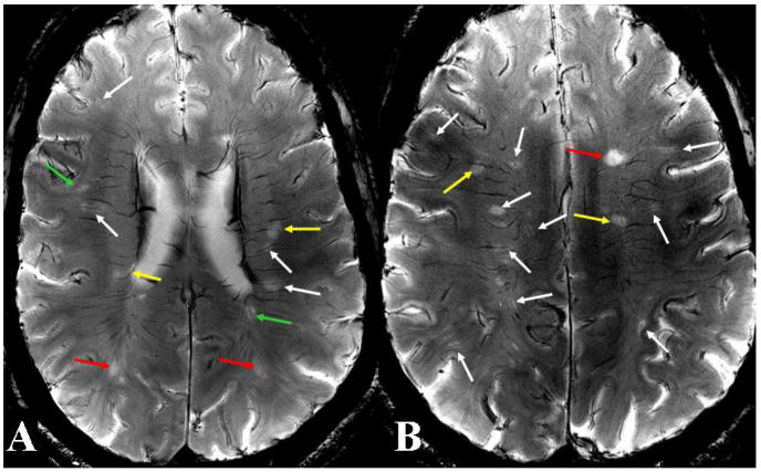
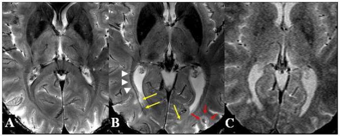
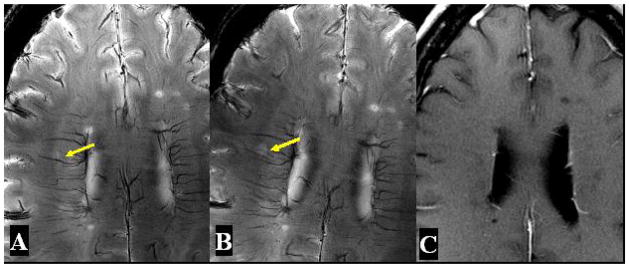
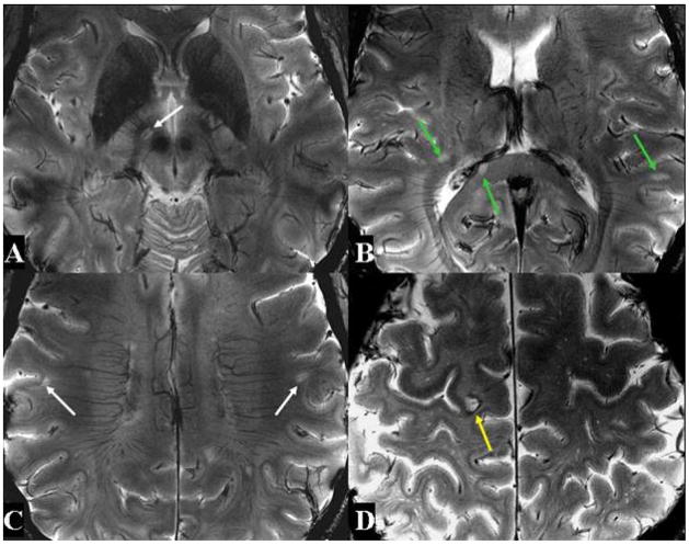
Results: We demonstrated markedly enhanced detection of unique microvascular involvement associated with most of the visualized MS lesions with abnormal signals on and around the venous wall on 7-T compared with 3-T MRI.
Conclusions: These findings, which have never been shown on conventional fields of MRI, not only allow for direct evidence of vascular pathogenesis in MS in vivo but also have important implications for monitoring lesion activity and therapeutic response.
Figures
Comment in
-
The retina as a window to the brain.Arch Neurol. 2008 Nov;65(11):1547-8; author reply 1548. doi: 10.1001/archneur.65.11.1547. Arch Neurol. 2008. PMID: 19001181 No abstract available.
References
-
- Rindficisch E. Histologisches detail zu der grauen degeneration von gehirn und ruckenmark. Archives of Pathological Anatomy and Physiology. 1863;26:474–483.
-
- Christoforidis GA, Bourekas EC, Baujan M, et al. High resolution MRI of the deep brain vascular anatomy at 8 Tesla: susceptibility-based enhancement of the venous structures. J Comput Assist Tomogr. 1999;23:857–66. - PubMed
-
- Poser CM, Paty DW, Scheinberg L, et al. New diagnostic criteria for multiple sclerosis: guidelines for research protocols. Ann Neurol. 1983;13:227–31. - PubMed
-
- Dawson J. The histology of disseminated sclerosis. Trans Roy Soc Edinb. 1916;50:517.
-
- Adams CW, Poston RN, Buk SJ. Pathology, histochemistry and immunocytochemistry of lesions in acute multiple sclerosis. J Neurol Sci. 1989;92:291–306. - PubMed
