. 2008 Jun;126(6):765-71.
doi: 10.1001/archopht.126.6.765.

Detailed visualization of the anterior segment using fourier-domain optical coherence tomography

Affiliations

Detailed visualization of the anterior segment using fourier-domain optical coherence tomography

Sanjay Asrani et al. Arch Ophthalmol. 2008 Jun.

Abstract

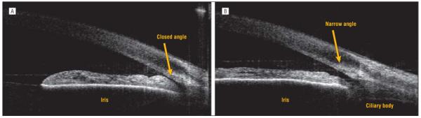
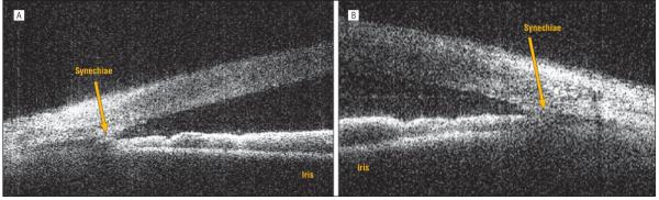
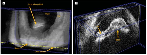
Objective: To study details of the anterior chamber drainage angle using Fourier-domain optical coherence tomography in healthy subjects and patients with angle abnormalities.

Methods: A high-speed anterior segment optical coherence tomography prototype was developed using a 1310-nm-wavelength swept light source. Six healthy subjects and 6 patients with glaucoma were imaged in an observational cross-sectional study.

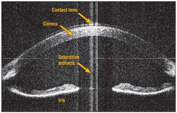
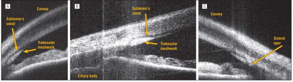
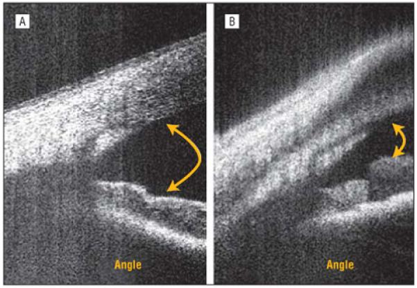
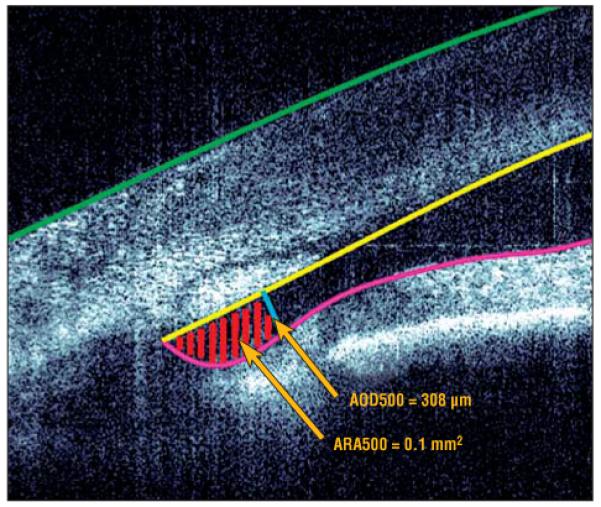
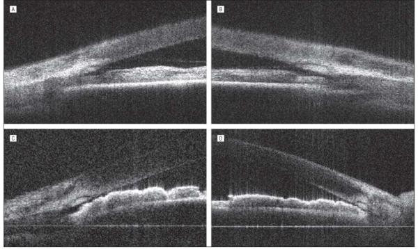
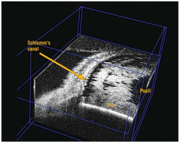
Results: Schlemm's canal and the trabecular meshwork were visualized in all of the patients. Fifteen-millimeter scans enabled entire anterior segment visualization providing configuration details of the iris with respect to the angle. Four-millimeter scans permitted detailed views of the angle configuration and its structures. Volumetric imaging was possible and Schlemm's canal was visualized along part of its circumference.

Conclusion: Anterior segment Fourier-domain optical coherence tomography permits detailed noncontact imaging of the angle and its structures, providing a tool to improve our understanding of the pathogenesis of narrow-angle glaucoma.


Figures

Figure 1
Figure 1
Concave iris configuration seen in a patient with mild myopia. The full depth of the anterior chamber can be visualized.
Figure 2
Figure 2
Detailed angle structures. A, The trabecular meshwork is visible as a triangular body and Schlemm’s canal as a curvilinear lucent space external to the trabecular meshwork. B, The ciliary body outline is visible along with Schlemm’s canal and the trabecular meshwork in another eye of a healthy subject. C, Note the scleral spur as a structure projecting into the anterior chamber at the root of the trabecular meshwork.
Figure 3
Figure 3
Angle configurations. A, An eye clinically open to the ciliary body band on gonioscopy. B, An eye open to the scleral spur on gonioscopy.
Figure 4
Figure 4
Representative narrow angle measurement. The epithelium, endothelium, and iris were manually delineated in unprocessed coordinates. The angle opening distance at the 500-μm (AOD500) line is shown in blue, and the angle recess area at the 500-μm (ARA500) region is hatched with red lines in unprocessed coordinates. The values for the AOD500 and ARA500 were calculated in processed coordinates, and the ARA500 was obtained by integration of the equations describing the lines.
Figure 5
Figure 5
High-resolution Fourier-domain optical coherence tomographic scans of narrow angles with gonioscopic grade I and grade 0. A, A very narrow space is visible between the root of the iris and the trabecular meshwork in the right eye of a patient with narrow-angle glaucoma (gonioscopic grade I). B, The left eye of the same patient (A) shows similar findings. C, Near touch between the trabecular meshwork and the root of the iris is visible in the right eye of another patient with narrow-angle glaucoma (gonioscopic grade 0). D, The left eye of the same patient (C) shows a similar angle configuration.
Figure 6
Figure 6
Images with the room lights turned off (A) and back on (B). A, A closed angle with the iris root almost in approximation with the trabecular meshwork when the room lights are off. B, On turning the room lights on, the pupil constricts with ensuing stretching of the iris and widening of the angle.
Figure 7
Figure 7
Peripheral anterior synechiae in a patient with narrow-angle glaucoma. A, Adhesion between the iris root and the trabecular meshwork is visible in the right eye. B, Similar findings are seen in the left eye of the same patient.
Figure 8
Figure 8
Cross-sectional planes of a volumetric image of a normal anterior segment showing Schlemm’s canal along part of the circumference by removing the overlying layers in the rendered 3-dimensional image.
Figure 9
Figure 9
An eye with a ciliary body tumor. A, A volumetric rendering showing irregularity of the anterior chamber depth due to elevation of the peripheral iris in the inferior region of the eye. B, A cross-sectional plane through the tumor location. Note that the actual tumor of the ciliary body is not visible.
Figure 10
Figure 10
Multiloculated iris cyst in the iris pigment epithelium.

References

    1. Huang D, Swanson EA, Lin CP, et al. Optical coherence tomography. Science. 1991;254(5035):1178–1181. - PMC - PubMed
    1. Radhakrishnan S, Goldsmith J, Huang D, et al. Comparison of optical coherence tomography and ultrasound biomicroscopy for detection of narrow anterior chamber angles. Arch Ophthalmol. 2005;123(8):1053–1059. - PubMed
    1. Radhakrishnan S, Huang D, Smith SD. Optical coherence tomography imaging of the anterior chamber angle. Ophthalmol Clin North Am. 2005;18(3):375–381. - PubMed
    1. Geerling G, Muller M, Winter C, et al. Intraoperative 2-dimensional optical coherence tomography as a new tool for anterior segment surgery. Arch Ophthalmol. 2005;123(2):253–257. - PubMed
    1. Leitgeb R, Hitzenberger CK, Fercher AF. Performance of fourier domain vs time domain optical coherence tomography. Opt Express. 2003;11(8):889–894. - PubMed

Publication types