

Retinal morphological changes of patients with X-linked retinoschisis evaluated by Fourier-domain optical coherence tomography
- PMID: 18541843
- PMCID: PMC2612690
- DOI: 10.1001/archopht.126.6.807
Retinal morphological changes of patients with X-linked retinoschisis evaluated by Fourier-domain optical coherence tomography
Abstract
Objective: To investigate the retinal microstructure and lamination of patients affected with X-linked retinoschisis (XLRS) using high-resolution imaging modalities.
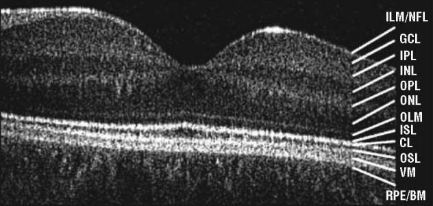
Methods: Patients diagnosed as having XLRS underwent assessment. Visual function testing included visual acuity, color vision, and full-field electroretinography. We used a high-resolution Fourier-domain optical coherence tomography (FD-OCT) system (4.5-mum axial resolution; 9 frames/s; 1000 A-scans per frame) combined with a handheld scanner. Macular image evaluation included schisis localization and retinal layer integrity.
Results: Six patients with XLRS and identified mutations in the XLRS1 gene underwent testing. Visual acuity ranged from 0.2 to 1.6 logMAR (logarithm of the minimum angle of resolution). Results of FD-OCT revealed foveal schisis extending from the outer to the inner plexiform layer in 4 of 6 patients. Bullous foveal schisis was associated with younger age. All patients showed extrafoveal schisis within the outer and inner nuclear and ganglion cell layer, alone or in combination. Photoreceptor outer and inner segment layers were disrupted and irregular in all patients.
Conclusions: Retinal dystrophy in XLRS is reflected by morphological changes within the inner and outer retinal layers. Disturbed foveal photoreceptor integrity was identified in all patients. Retinal layer abnormalities correlated with age but did not appear to correlate with visual acuity or genotypic variation.
Figures

References
-
- Haas J. Ueber das Zusammenvorkommen von Veraenderungen der Retina und Chorioidea. Archive Augenheilkunde. 1898;37:343–348.
-
- Tantri A, Vrabec TR, Cu-Unjieng A, Frost A, Annesley WH, Jr, Donoso LA. X-linked retinoschisis: a clinical and molecular genetic review. Surv Ophthalmol. 2004;49(2):214–230. - PubMed
-
- Apushkin MA, Fishman GA, Janowicz MJ. Correlation of optical coherence tomography findings with visual acuity and macular lesions in patients with X-linked retinoschisis. Ophthalmology. 2005;112(3):495–501. - PubMed
-
- Sauer CG, Gehrig A, Warneke-Wittstock R, et al. Positional cloning of the gene associated with X-linked juvenile retinoschisis. Nat Genet. 1997;17(2):164–170. - PubMed
-
- Molday LL, Hicks D, Sauer CG, Weber BH, Molday RS. Expression of X-linked retinoschisis protein RS1 in photoreceptor and bipolar cells. Invest Ophthalmol Vis Sci. 2001;42(3):816–825. - PubMed
Publication types
MeSH terms
Substances
Grants and funding
LinkOut - more resources
Full Text Sources
Other Literature Sources
