. 2008 Sep;466(9):2081-91.
doi: 10.1007/s11999-008-0327-z. Epub 2008 Jun 10.

Histogenetic characterization of giant cell tumor of bone

Affiliations

Histogenetic characterization of giant cell tumor of bone

Manuela Salerno et al. Clin Orthop Relat Res. 2008 Sep.

Abstract

The unpredictable behavior of giant cell tumor (GCT) parallels its controversial histogenesis. Multinucleated giant cells, stromal cells, and CD68(+) monocytes/macrophages are the three elements that interact in GCT. We compared the ability of stromal cells and normal mesenchymal stromal cells to differentiate into osteoblasts. Stromal cells and mesenchymal cells had similar proliferation rates and lifespans. Although stromal cells expressed early osteogenic markers, they were unable to differentiate into osteoblasts but they did express intracellular adhesion molecule-1, a marker of bone-lining cells. They were unable to form clones in a semisolid medium and unable to promote osteoclast differentiation, although they exerted a strong chemotactic effect on osteoclast precursors. Stromal cells may be either immature proliferating osteogenic elements or specialized osteoblast-like cells that fail to show neoplastic features but can induce the differentiation of osteoclast precursors. They might be secondarily induced to proliferate by a paracrine effect induced by monocyte-macrophages and/or giant cells. The increased number of giant cells in GCT may be secondary to an autocrine circuit mediated by the receptor activator of nuclear factor kB.


Figures
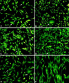
Fig. 1A–F
Fig. 1A–F
In vitro morphologic features of stromal cells (SC) and human mesenchymal stromal cells (hMSC) are shown. Cell morphology was assessed in SC-1 (A), SC-2 (B), SC-3 (C), SC-4 (D), SC-5 (E), and hMSC (F) after acridine orange staining of adherent cells. SC cultures were composed of spindle-shaped and polygonal (arrows) cells, whereas hMSC were homogeneous cultures of flattened cells. In SC cultures, binuclear cells were also observed (stain, acridine orange; original magnification, x10; bar = 50 μm).
Fig. 2A–E
Fig. 2A–E
The expression of osteoblastic lineage-related genes is shown. The mRNA expression of osteoblast-related genes was analyzed in giant cell tumor (GCT) tissues, in untreated stromal cells (SC), or in human mesenchymal stromal cells (hMSC) (T0) and in SC or in hMSC after 14 days in differentiating medium (T1) (hMSC, n = 3). Gel electrophoresis results for GCT tissues (A); gel electrophoresis results and semiquantitative analysis for SC and hMSC for core binding factor a-1 (Cbfa1) (B), osteocalcin (C), Type I collagen (D), and intracellular adhesion molecule-1 (E). In the upper panel, a representative image of the amplification product is shown, whereas in the lower panel, specific bands were quantified by dedicated software for densitometric evaluation. Each amplified product of the corresponding gene was normalized to β-actin signals determined in parallel for each sample (BE); otherwise, only a representative image of the amplification product as a single panel is shown (A).
Fig. 3A–C
Fig. 3A–C
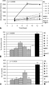
Proliferation rate and colony-forming unit (CFU) efficiency of stromal cells (SC) are shown. Cell proliferation, Ki67 index, and clonal efficiency were evaluated in SC and compared with human mesenchymal stromal cells (hMSC) or Saos-2 cells. (A) 3-(4,5-dimethylthiazol-2-yl)-2,5-diphenyltetrazolium assay (hMSC n = 2) (A); Ki67 index (hMSC n = 2) (B); number of colonies formed after 1 to 2 weeks (hMSC n = 3) (C). The proliferation rate of SC is comparable to that of hMSC, whereas CFU is higher.
Fig. 4A–B
Fig. 4A–B
Chemotaxis of CD14+-monocyte induced by stromal cells (SC) is shown. The ability of SC to chemoattract CD14+ human blood monocytes and the mRNA expression of the chemoattractive factor stromal cell derived factor-1 (SDF-1) were compared with those of human mesenchymal stromal cells (hMSC). Number of migrated cells after coculture with SC or hMSC (n = 3) (A); gel electrophoresis results for SDF-1 in SC, hMSC, and in giant cell tumor tissues (B). SC induced considerable migration of CD14+ monocytes, although this ability was not dependent on SDF-1 production.
Fig. 5A–C
Fig. 5A–C
Osteoclast differentiation and activity induced by stromal cells (SC) are shown. The isoform 5b of tartrate-resistant acid phosphatase (TRACP) and collagen resorption activity were evaluated in coculture of peripheral blood monocytes with SC and human mesenchymal stromal cells (hMSC). The expression of receptor activator of nuclear factor kB (RANKL) mRNA was also analyzed in SC, hMSC, and giant cell tumor tissues (hMSC n = 2). For TRACP 5b assay (A), and Osteolyse assay (B), p was calculated for the difference between SC and hMSC. (C) Gel electrophoresis results for RANKL. SC did not induce osteoclast activity or differentiation.
Fig. 6A–B
Fig. 6A–B
Histogenesis of giant cell tumor (GCT) is shown. Hypothetical models of interaction between stromal cells (SC) and giant cells (GC) in GCT. Neoplastic osteoblast-like stromal cells induce osteoclast activation and differentiation through receptor activator of nuclear factor kB (RANKL) secretion (A). Multinucleated GC/monocytes promote osteoclast activation and differentiation as a result of an autocrine RANKL-mediated circuit, in turn inducing hyperplastic proliferation and activation of osteoblast-like intracellular adhesion molecule-1-positive “bone lining cells” (B).

References

    1. {'text': '', 'ref_index': 1, 'ids': [{'type': 'DOI', 'value': '10.1002/path.1711470302', 'is_inner': False, 'url': 'https://doi.org/10.1002/path.1711470302'}, {'type': 'PubMed', 'value': '4067733', 'is_inner': True, 'url': 'https://pubmed.ncbi.nlm.nih.gov/4067733/'}]}
    2. Athanasou NA, Bliss E, Gatter KC, Heryet A, Woods CG, McGee JO. An immunohistological study of giant-cell tumour of bone: evidence for an osteoclast origin of the giant cells. J Pathol. 1985;147:153–158. - PubMed
    1. {'text': '', 'ref_index': 1, 'ids': [{'type': 'PubMed', 'value': '7285463', 'is_inner': True, 'url': 'https://pubmed.ncbi.nlm.nih.gov/7285463/'}]}
    2. Bauer FC, Urist MR. The reaction of giant cell tumors of bone to transplantation into athmic mice: including observations on osteoinduction in the host bed. Clin Orthop Relat Res. 1981;159:257–264. - PubMed
    1. {'text': '', 'ref_index': 1, 'ids': [{'type': 'PubMed', 'value': '3056645', 'is_inner': True, 'url': 'https://pubmed.ncbi.nlm.nih.gov/3056645/'}]}
    2. Bertoni F, Present D, Sudanese A, Baldini N, Bacchini P, Campanacci M. Giant-cell tumor of bone with pulmonary metastases: six case reports and a review of the literature. Clin Orthop Relat Res. 1988;237:275–285. - PubMed
    1. {'text': '', 'ref_index': 1, 'ids': [{'type': 'DOI', 'value': '10.1126/science.279.5349.349', 'is_inner': False, 'url': 'https://doi.org/10.1126/science.279.5349.349'}, {'type': 'PubMed', 'value': '9454332', 'is_inner': True, 'url': 'https://pubmed.ncbi.nlm.nih.gov/9454332/'}]}
    2. Bodnar AG, Ouellette M, Frolkis M, Holt SE, Chiu CP, Morin GB, Harley CB, Shay JW, Lichtsteiner S, Wright WE. Extension of life-span by introduction of telomerase into normal human cells. Science. 1998;279:349–352. - PubMed
    1. {'text': '', 'ref_index': 1, 'ids': [{'type': 'DOI', 'value': '10.1007/s002560050230', 'is_inner': False, 'url': 'https://doi.org/10.1007/s002560050230'}, {'type': 'PubMed', 'value': '9151375', 'is_inner': True, 'url': 'https://pubmed.ncbi.nlm.nih.gov/9151375/'}]}
    2. Brien EW, Mirra JM, Kessler S, Suen M, Ho JK, Yang WT. Benign giant cell tumor of bone with osteosarcomatous transformation (‘dedifferentiated’ primary malignant GCT): report of two cases. Skeletal Radiol. 1997;26:246–255. - PubMed

Publication types