Review
. 2008 Sep;60(9):609-19.
doi: 10.1002/iub.98.

Glutamate forward and reverse transport: from molecular mechanism to transporter-mediated release after ischemia

Affiliations
Review

Glutamate forward and reverse transport: from molecular mechanism to transporter-mediated release after ischemia

Christof Grewer et al. IUBMB Life. 2008 Sep.

Abstract

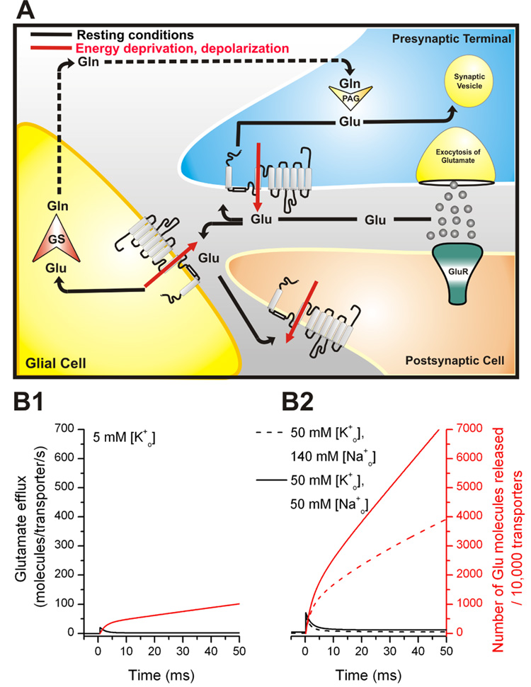
Glutamate transporters remove the excitatory neurotransmitter glutamate from the extracellular space after neurotransmission is complete, by taking glutamate up into neurons and glia cells. As thermodynamic machines, these transporters can also run in reverse, releasing glutamate into the extracellular space. Because glutamate is excitotoxic, this transporter-mediated release is detrimental to the health of neurons and axons, and it, thus, contributes to the brain damage that typically follows a stroke. This review highlights current ideas about the molecular mechanisms underlying glutamate uptake and glutamate reverse transport. It also discusses the implications of transporter-mediated glutamate release for cellular function under physiological and patho-physiological conditions.


Figures

Figure 1
Figure 1
(A) Illustration of the stoichiometry of glutamate transporters (left panel). The plus sign (+) inside the cell indicates depolarization of the cell induced by inward glutamate transport. The right panel illustrates the glutamate-induced anion conductance. (B) Top view of the trimeric assembly of GltPh adapted from (32). (C) Transmembrane topology of glutamate transporters. The two Tl+ (thallium+) binding sites found in GltPh are highlighted in blue, the bound substrate is shown in green. The functionally important C-terminal peptide is highlighted in red.
Figure 2
Figure 2
(A) Proposed mechanism for the transmembrane movement of glutamate catalyzed by the glutamate transporter, based on functional and structural data. (B) Pre-steady-state currents measured in response to a rapid glutamate concentration jump point to at least two independent processes associated with glutamate translocation. (C) The temperature dependence of pre-steady-state kinetics indicates the existence of two conformational changes.
Figure 3
Figure 3
Reverse translocation of glutamate is associated with transmembrane charge movement. (A) Illustration of the rapid delivery of glutamate to the cytosolic face of the membrane by laser photolysis of intracellular caged glutamate. (B) and (C) show reverse transport currents induced by photolysis of 6 mM MNI-Glu at t = 0 ms (yellow arrow) in the reverse transport mode (B, illustrated in top panel) and reverse exchange mode (C, illustrated in top panel).
Figure 4
Figure 4
A simple kinetic model based on a “first-in-first-out” mechanism can explain the experimental data (A). (B1) and (B2) illustrate possible substrate/Na+ association/dissociation sequences based on mirror-symmetry and glide symmetry models.
Figure 5
Figure 5
(A) Distribution and function of EAA transporters at the synapse during a synaptic event. Glutamate (circles) binding to glutamate receptors (GluR) mediates the signal to postsynaptic neurons. Rapid removal of glutamate from the synaptic cleft is achieved by glutamate transporters which are located in glial cells and neurons. Once glutamate is transported into the cell (indicated by arrows), it can be sequestered in synaptic vesicles, ready for a new cycle of neurotransmission (indicated by arrows in the presynapse) or is converted in glial cells by the glia-specific enzyme glutamine-synthetase (GS) to glutamine (Gln), which is released into the extracellular space, and subsequently taken up by glutamatergic neurons for a new cycle of transmitter synthesis (indicated by dotted lines) for a new transmitter synthesis. The red arrows indicate transporter-mediated glutamate release upon energy deprivation and/or membrane depolarization. (B1) Predicted rate of glutamate efflux (black lines) in response to a step depolarization from −80 mV to −40 mV at t = 0 ms in the presence of 5 mM extracellular [K+] (physiological case, left Y-axis) (B2) Predicted rate of glutamate efflux (black lines) in response to a step depolarization from −80 mV to −40 mV at t = 0 ms in the presence of 50 mM extracellular [K+] (normal [Na+]o, dashed lines) and 50 mM extracellular [K+] (reduced [Na+]o of 50 mM, solid lines, pathophysiological case). The red lines show the total number of glutamate molecules released by 10,000 transporters (right Y-axis).

References

    1. Danbolt NC. Glutamate uptake. Prog. Neurobiol. 2001;65:101–105. - PubMed
    1. Grewer C, Rauen T. Electrogenic Glutamate Transporters in the CNS: Molecular Mechanism, Pre-steady-state Kinetics, and their Impact on Synaptic Signaling. J. Membr. Biol. 2005;203:1–20. - PMC - PubMed
    1. Sheldon AL, Robinson MB. The role of glutamate transporters in neurodegenerative diseases and potential opportunities for intervention. Neurochem. Int. 2007;51:333–355. - PMC - PubMed
    1. Vandenberg RJ. Molecular pharmacology and physiology of glutamate transporters in the central nervous system. Clin. Exp. Pharmacol. Physiol. 1998;25:393–400. - PubMed
    1. Kanai Y, Hediger MA. The glutamate and neutral amino acid transporter family: physiological and pharmacological implications. Eur. J. Pharmacol. 2003;479:237–247. - PubMed

Publication types

MeSH terms

Substances