

Losartan reduced connexin43 expression in left ventricular myocardium of spontaneously hypertensive rats
- PMID: 18543397
- PMCID: PMC2408697
- DOI: 10.1631/jzus.B0820050
Losartan reduced connexin43 expression in left ventricular myocardium of spontaneously hypertensive rats
Abstract
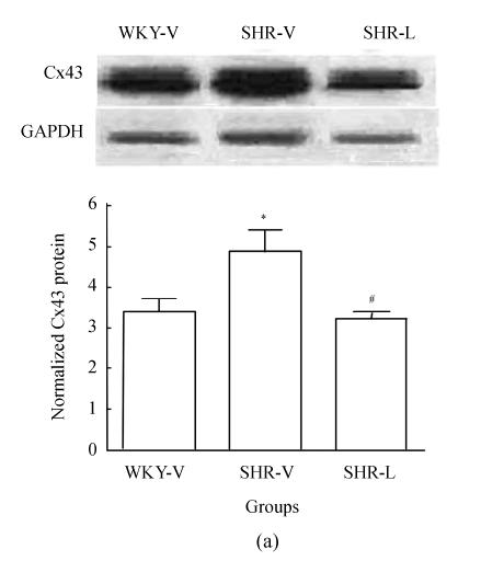
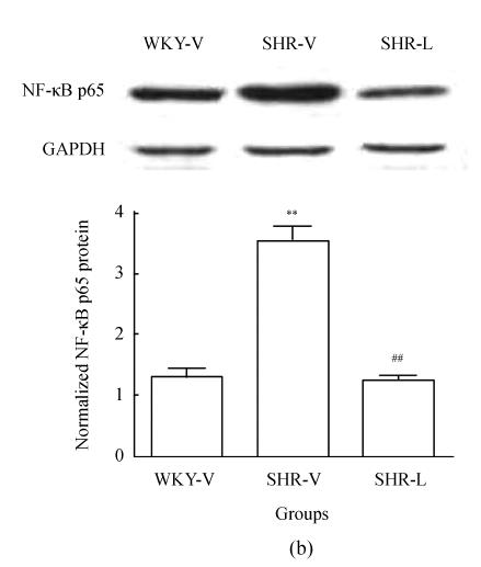
Objective: To assess the effect of angiotensin II type 1 (AT(1)) receptor antagonist losartan on myocardium connexin43 (Cx43) gap junction (GJ) expression in spontaneously hypertensive rats (SHRs) and investigate possible mechanisms.
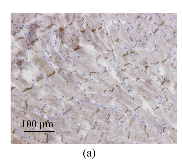
Methods: Sixteen 9-week-old male SHRs and 8 age-matched male Wistar-Kyoto (WKY) rats were included in this study. SHRs were randomly divided into two groups to receive losartan at 30 mg/(kg x d) by oral gavage once daily for 8 weeks (SHR-L) or vehicle (0.9% saline) to act as controls (SHR-V); WKY rats receiving vehicle for 8 weeks served as normotensive controls. At the end of the experiment, rats were sacrificed and the hearts were removed. Expressions of Cx43 and nuclear factor-kappaB p65 (NF-kappaB p65) proteins in all three groups were observed and further investigations on the effect of angiotensin II type 1 receptor antagonist losartan (30 mg/(kg x d), 8 weeks) on Cx43 expression were conducted with Western blot and immunohistochemistry. NF-kappaB p65 protein in nuclear extracts was determined by Western blot.
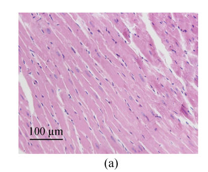
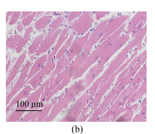
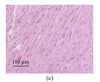
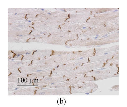
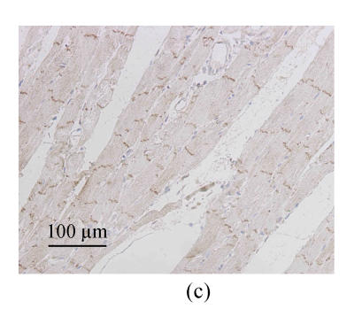
Results: Left ventricular (LV) hypertrophy was prominent in SHRs, Cx43 and NF-kappaB p65 protein expressions were obviously upregulated and Cx43 distribution was dispersed over the cell surface. Treatment with losarton reduced the over-expressions of Cx43 and NF-kappaB p65 in LV myocardium. The distribution of Cx43 gap junction also became much regular and confined to intercalated disk after losartan treatment.
Conclusion: Cx43 level was upregulated in LV myocardium of SHR during early stage of hypertrophy. Angiotensin II type 1 receptor antagonist losartan prevented Cx43 gap junction remodeling in hypertrophied left ventricles, possibly through the NF-kappaB pathway.
Figures
Similar articles
-
Atorvastatin prevents connexin43 remodeling in hypertrophied left ventricular myocardium of spontaneously hypertensive rats.Chin Med J (Engl). 2007 Nov 5;120(21):1902-7. Chin Med J (Engl). 2007. PMID: 18067764
-
[PPAR alpha activator fenofibrate regressed left ventricular hypertrophy and increased myocardium PPAR alpha expression in spontaneously hypertensive rats].Zhejiang Da Xue Xue Bao Yi Xue Ban. 2007 Sep;36(5):470-6. doi: 10.3785/j.issn.1008-9292.2007.05.011. Zhejiang Da Xue Xue Bao Yi Xue Ban. 2007. PMID: 17924466 Chinese.
-
Transient prehypertensive treatment in spontaneously hypertensive rats: a comparison of losartan and amlodipine regarding long-term blood pressure, cardiac and renal protection.Int J Mol Med. 2012 Dec;30(6):1376-86. doi: 10.3892/ijmm.2012.1153. Epub 2012 Oct 9. Int J Mol Med. 2012. PMID: 23064712
-
Losartan inhibits the post-transcriptional synthesis of collagen type I and reverses left ventricular fibrosis in spontaneously hypertensive rats.J Hypertens. 1999 Jan;17(1):107-14. doi: 10.1097/00004872-199917010-00016. J Hypertens. 1999. PMID: 10100101
-
Chronic AT(1) blockade stimulates extracellular collagen type I degradation and reverses myocardial fibrosis in spontaneously hypertensive rats.Hypertension. 2000 Jun;35(6):1197-202. doi: 10.1161/01.hyp.35.6.1197. Hypertension. 2000. PMID: 10856263
Cited by
-
Changes of gene expressions in spontaneously hypertensive rat model after losartan treatment.Korean Circ J. 2012 Nov;42(11):761-8. doi: 10.4070/kcj.2012.42.11.761. Epub 2012 Nov 28. Korean Circ J. 2012. PMID: 23236328 Free PMC article.
-
The therapeutic effect of rosuvastatin on cardiac remodelling from hypertrophy to fibrosis during the end-stage hypertension in rats.J Cell Mol Med. 2012 Sep;16(9):2227-37. doi: 10.1111/j.1582-4934.2012.01536.x. J Cell Mol Med. 2012. PMID: 22288611 Free PMC article.
-
Connexin 43 is an emerging therapeutic target in ischemia/reperfusion injury, cardioprotection and neuroprotection.Pharmacol Ther. 2015 Sep;153:90-106. doi: 10.1016/j.pharmthera.2015.06.005. Epub 2015 Jun 11. Pharmacol Ther. 2015. PMID: 26073311 Free PMC article. Review.
-
Connexins in Cardiovascular and Neurovascular Health and Disease: Pharmacological Implications.Pharmacol Rev. 2017 Oct;69(4):396-478. doi: 10.1124/pr.115.012062. Pharmacol Rev. 2017. PMID: 28931622 Free PMC article. Review.
-
Aliskiren-attenuated myocardium apoptosis via regulation of autophagy and connexin-43 in aged spontaneously hypertensive rats.J Cell Mol Med. 2014 Jul;18(7):1247-56. doi: 10.1111/jcmm.12273. Epub 2014 Apr 6. J Cell Mol Med. 2014. PMID: 24702827 Free PMC article.
References
-
- Dahlof B, Devereux RB, Kjeldsen SE, Julius S, Beevers G, de Faire U, Fyhrquist F, Ibsen H, Kristiansson K. Cardiovascular morbidity and mortality in the losartan intervention for endpoint reduction in hypertension study (LIFE): a randomized trial against atenolol. Lancet. 2002;359(9311):995–1003. doi: 10.1016/S0140-6736(02)08089-3. - DOI - PubMed
-
- Emdad L, Uzzaman M, Takagshi Y, Honjo H, Uchida T, Severs NJ, Kodama I, Murata Y. Gap junction remodeling in hypertrophied left ventricles of aortic-banded rats: prevention by angiotensin II type 1 receptor blockade. J Mol Cell Cardiol. 2001;33(2):219–231. doi: 10.1006/jmcc.2000.1293. - DOI - PubMed
MeSH terms
Substances
LinkOut - more resources
Full Text Sources
Medical
Molecular Biology Databases
Research Materials
Miscellaneous
