Review
. 2008:267:183-206.
doi: 10.1016/S1937-6448(08)00604-7.

New insights into mechanism and regulation of actin capping protein

Affiliations
Review

New insights into mechanism and regulation of actin capping protein

John A Cooper et al. Int Rev Cell Mol Biol. 2008.

Abstract

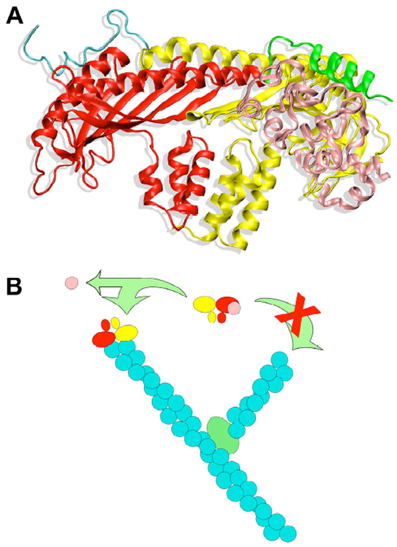
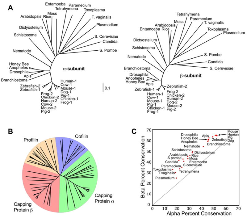
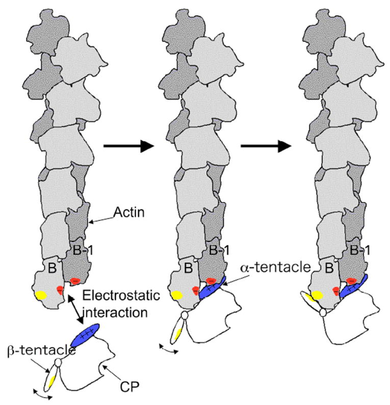
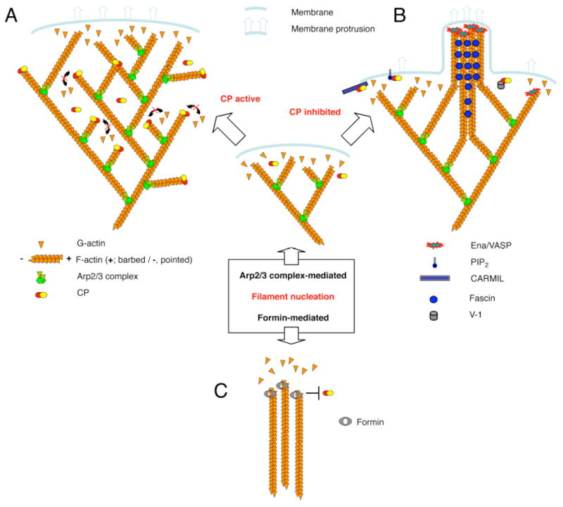
The heterodimeric actin capping protein, referred to here as "CP," is an essential element of the actin cytoskeleton, binding to the barbed ends of actin filaments and regulating their polymerization. In vitro, CP has a critical role in the dendritic nucleation process of actin assembly mediated by Arp2/3 complex, and in vivo, CP is important for actin assembly and actin-based process of morphogenesis and differentiation. Recent studies have provided new insight into the mechanism of CP binding the barbed end, which raises new possibilities for the dynamics of CP and actin in cells. In addition, a number of molecules that bind and regulate CP have been discovered, suggesting new ideas for how CP may integrate into diverse processes of cell physiology.


Figures

Figure 1
Figure 1
Illustrations of a model for the interaction of V-1 with CP and how their interaction inhibits actin capping activity. A) The structure of the proposed molecular interaction between capping protein and V-1/ myotrophin. The α subunit of capping protein is yellow, and its C-terminal actin-binding region is teal. The β subunit of capping protein is red, and its C-terminal actin-binding region is green. V-1 / myotrophin is in pink. B) The binding of V-1/myotrophin prevents capping protein from binding to actin filament barbed ends, i.e. “capping.” Growth of free barbed ends is an essential of the dendritic nucleation model for actin assembly, which involves Arp2/3 complex as the nucleating and branching agent. The color scheme is similar to the one in panel A, with Arp2/3 complex as the green oval at an end-to-side branch point for two actin filaments, whose subunits are teal.
Figure 2
Figure 2
Phylogenetic analysis of CP subunits. Amino acid sequences were identified in a wide range of eukaryotes by BLAST. The sequences were aligned with CLUSTALW. A) Phylogenetic trees for the α and β subunits are remarkably similar. Vertebrates have up to three isoforms of each subunit, while invertebrates and lower organisms have single isoforms of each subunit. Vertebrate isoforms 1 and 2 represent nearly all the CP outside of germ cells, they cluster into distinct groups. B) Phylogenetic analysis of CP subunits compared with cofilin and profilin, other actin-binding proteins. The α and β subunits of CP are not more similar to each other than they are to the cofilin and profilin families, despite their similar secondary structures and interactions with actin. C) For each organism, the similarity of its β subunit to the β subunits of other organisms is plotted versus the similarity of its α subunit to the α subunits of other organisms. For vertebrates, only a single isoform of each subunit was included per species. The results show that the β subunit sequences are more similar to each other than are those of the α subunits.
Figure 3
Figure 3
A model for the binding of CP to the actin filament barbed-end proposed by Narita and colleagues (Narita et al., 2006), kindly provided by the authors and reproduced with their permission. Basic residues on the CP α C-terminal region /(blue) are attracted to acidic residues on the barbed end of the actin filament (red). These acidic residues include ones from the terminal and the penultimate protomers of the filament, labeled B and B-1. Next, the mobile β tentacle searches for its binding position on the filament. Finally, the hydrophobic surface (yellow) of the amphipathic β tentacle binds to the hydrophobic cleft (yellow) on the terminal protomer, B.
Figure 4
Figure 4
Illustration of potential modes of actin assembly in cells with respect to CP, based on one created and provided by Dr. Martin Wear (Wear and Cooper, 2004b). A) When CP is active and actin nucleation is Arp2/3-mediated, lamellipodial assembly predominates. Newly created free barbed ends are near the membrane. They elongate to push the membrane forward and / or the actin filament network backward. After some time, CP caps those barbed ends, which would seem to be efficacious because the ends are no longer near the membrane and their further growth would not produce useful work. B) In this setting, when CP is inactivated in one location, by any of several potential inhibitors, then the filaments in that small region may continue to grow, producing a thin protrusion that contains a bundle of actin filaments. C) An alternative mechanism that may produce actin filament bundles, perhaps not associated with a plasma membrane, is the nucleation of actin polymerization by a formin. Formins allow actin subunits to add and do not allow CP to add. Thus, the filaments continue to grow.
Figure 5
Figure 5
An illustration of a new model for the structure of the Z-disc, provided by Dr. Carol Gregorio. Based on a structure / function analysis of the interaction of nebulin with CapZ (Pappas et al., 2007), the model proposes that the nebulin molecule crosses from one actin-based thin filament to another one, within the Z-disc.

Similar articles

Cited by

References

    1. Applewhite DA, Barzik M, Kojima S, Svitkina TM, Gertler FB, Borisy GG. Ena/VASP proteins have an anti-capping independent function in filopodia formation. Mol Biol Cell. 2007;18:2579–2591. - PMC - PubMed
    1. Barkalow K, Witke W, Kwiatkowski DJ, Hartwig JH. Coordinated regulation of platelet actin filament barbed ends by gelsolin and capping protein. J Cell Biol. 1996;2:389–399. - PMC - PubMed
    1. Barzik M, Kotova TI, Higgs HN, Hazelwood L, Hanein D, Gertler FB, Schafer DA. Ena/VASP proteins enhance actin polymerization in the presence of barbed end capping proteins. J Biol Chem. 2005;280:28653–28662. - PMC - PubMed
    1. Bearer EL. Direct observation of actin filament severing by gelsolin and binding by gCap39 and CapZ. J Cell Biol. 1991;115:1629–1638. - PMC - PubMed
    1. Bhattacharya N, Ghosh S, Sept D, Cooper JA. Binding of Myotrophin/V-1 to Actin-capping Protein: Implications for How Capping Protein binds to the Filament Barbed End. J Biol Chem. 2006;281:31021–31030. - PMC - PubMed

Publication types

MeSH terms

LinkOut - more resources