. 2008 Aug;29(8):1538-45.
doi: 10.1093/carcin/bgn137. Epub 2008 Jun 9.

Superoxide-mediated proteasomal degradation of Bcl-2 determines cell susceptibility to Cr(VI)-induced apoptosis

Affiliations

Superoxide-mediated proteasomal degradation of Bcl-2 determines cell susceptibility to Cr(VI)-induced apoptosis

Neelam Azad et al. Carcinogenesis. 2008 Aug.

Abstract

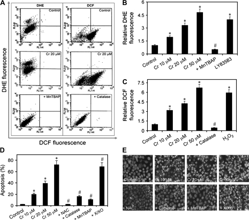
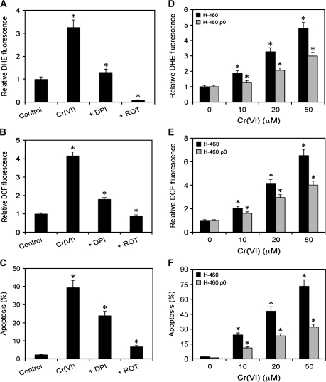
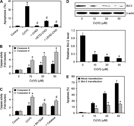
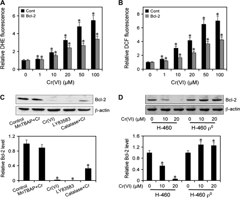
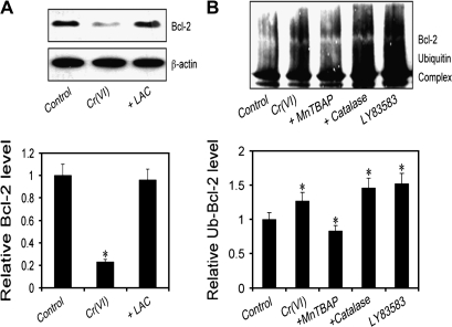
Hexavalent chromium [Cr(VI)] compounds are redox cycling environmental carcinogens that induce apoptosis as the primary mode of cell death. Defects in apoptosis regulatory mechanisms contribute to carcinogenesis induced by Cr(VI). Activation of apoptosis signaling pathways is tightly linked with the generation of reactive oxygen species (ROS). Likewise, ROS have been implicated in the regulation of Cr(VI)-induced apoptosis and carcinogenicity; however, its role in Cr(VI)-induced apoptosis and the underlying mechanism are largely unknown. We report that ROS, specifically superoxide anion (.O(-)(2), mediates Cr(VI)-induced apoptosis of human lung epithelial H460 cells. H460 rho(0) cells that lack mitochondrial DNA demonstrated a significant decrease in ROS production and apoptotic response to Cr(VI), indicating the involvement of mitochondrial ROS in Cr(VI)-induced apoptosis. In agreement with this observation, we found that Cr(VI) induces apoptosis mainly through the mitochondrial death pathway via caspase-9 activation, which is negatively regulated by the antiapoptotic protein Bcl-2. Furthermore, .O(-)(2) induced apoptosis in response to Cr(VI) exposure by downregulating and degrading Bcl-2 protein through the ubiquitin-proteasomal pathway. This study reveals a novel mechanism linking .O(-)(2) with Bcl-2 stability and provides a new dimension to ROS-mediated Bcl-2 downregulation and apoptosis induction.


Figures

Fig. 1.
Fig. 1.
Effects of Cr(VI) and ROS modulators on ROS levels and apoptosis in human lung epithelial H460 cells. (A, B and C) Subconfluent (90%) monolayers of H460 cells were treated with varying doses of Cr(VI) (0–50 μM) or were pretreated with MnTBAP (100 μM) or catalase (1000 U/μl) for 0.5 h followed by Cr(VI) (20 μM) treatment and were analyzed for ·O2 and H2O2 production by flow cytometry using DHE and DCF diacetate fluorescent probes, respectively. Plots show relative DHE or DCF fluorescence intensity over non-treated control determined at the peak response time of 1 h after Cr(VI) treatment. LY83583 (10 μM) and H2O2 (0.1 mM) were used as positive controls for DHE and DCF measurements, respectively. (D) Cells were exposed to Cr(VI) (0–50 μM) for 12 h or pretreated with MnTBAP (100 μM), catalase (1000 U/μl), N-acetyl cysteine (NAC) (10 mM) or xanthine/xanthine oxidase (X/XO) (1 μM/2 mU/ml) for 1 h followed by Cr(VI) (20 μM) treatment for 12 h and analyzed for apoptosis by Hoechst 33342 assay. (E) Fluorescence micrographs of treated cells stained with the Hoechst dye. Apoptotic cells exhibited shrunken and fragmented nuclei with bright nuclear fluorescence. Values are mean ± SD (n ≥ 3). *P < 0.05 versus non-treated control, #P < 0.05 versus 20 μM Cr(VI)-treated control.
Fig. 2.
Fig. 2.
Effects of GPx and SOD overexpression on Cr(VI)-induced apoptosis and ROS generation. (A) H460 cells were stably transfected with GPx, SOD or control pcDNA3 plasmid (Cont) as described under Materials and Methods. Cell lysates were analyzed for GPx and SOD expression by western blotting. β-Actin was used as a loading control. (B) Transfected cells were treated with Cr(VI) (1–100 μM) for 12 h and analyzed for apoptosis by Hoechst 33342 assay. (C) GPx- and mock-transfected cells were treated with Cr(VI) (0–100 μM) and analyzed for DCF fluorescence at 1 h posttreatment. (D) SOD- and mock-transfected cells were treated with Cr(VI) (0–100 μM) and analyzed for DHE fluorescence at 1 h posttreatment. Plots show relative fluorescence intensity over non-treated control. Values are mean ± SD (n ≥ 3).
Fig. 3.
Fig. 3.
Cellular source of Cr(VI)-induced ROS generation. (A and B) Subconfluent (90%) monolayers of H460 cells were either left untreated or pretreated with 5 μM of DPI or ROT for 0.5 h, followed by Cr(VI) treatment (20 μM) and analyzed for DHE and DCF fluorescence intensities after 1 h. (C) Cells were similarly treated with Cr(VI) and DPI or ROT and analyzed for apoptosis after 12 h by Hoechst assay. (D and E) H460 and H460 ρ0 cells were treated with Cr(VI) (0–50 μM) for 1 h and analyzed for DHE and DCF fluorescence intensities. Plots show relative fluorescence intensity over non-treated control. (F) Cells were similarly treated and analyzed for apoptosis after 12 h. Values are mean ± SD (n ≥ 3). *P < 0.05 versus non-treated control.
Fig. 4.
Fig. 4.
Caspase activation and Bcl-2 expression in response to Cr(VI). (A) Subconfluent (90%) monolayer of H460 cells were either left untreated or pretreated with zVAD-FMK (10 μM), IETD-CHO (2 μM) or LEHD-CHO (2 μM) for 1 h followed by Cr(VI) treatment (20 μM) and analyzed for apoptosis after 12 h. (B) Caspase activity assays of cells treated with Cr(VI) (0–50 μM) for 12 h. Cell lysates (20 μg protein) were prepared and analyzed for caspase-8 and -9 activity using specific fluorescent substrates IETD-AMC and LEHD-AMC, respectively. (C) Subconfluent (90%) monolayer of H460 cells were either left untreated or pretreated with MnTBAP (100 μM) or catalase (1000 U/ml) for 1 h followed by Cr(VI) treatment (20 μM) for 12 h. Caspase activity was measured as mentioned above. (D) Dose effect of Cr(VI) on Bcl-2 expression. Cells were treated with varying doses of Cr(VI) (0–50 μM) for 12 h and cell lysates were prepared and analyzed for Bcl-2 expression by western blotting. Blots were reprobed with β-actin antibody to confirm equal loading of samples. Immunoblot signals were quantified by densitometry and mean data from independent experiments were normalized to the result obtained in non-treated cells (control). (E) H460 cells were stably transfected with Bcl-2 or control pcDNA3 plasmid. Transfected cells were treated with Cr(VI) (0–50 μM) for 12 h and analyzed for apoptosis. Plots are mean ± SD (n = 4). *P < 0.05 versus non-treated or mock-transfected controls. #P < 0.05 versus 20 μM Cr(VI)-treated control.
Fig. 5.
Fig. 5.
Correlation between Cr(VI)-induced ROS generation and Bcl-2 expression. (A) and (B) H460 cells were stably transfected with Bcl-2 or control pcDNA3 plasmid. Transfected cells were treated with Cr(VI) (0–100 μM) for 1 h and analyzed for DHE and DCF fluorescence intensities. Plots show relative fluorescence intensity over non-treated control. (C) H460 cells were either left untreated or pretreated with MnTBAP (100 μM) or catalase (1000 U/μl) for 1 h and then treated with Cr(VI) (20 μM) for 12 h. Cells were also treated with LY83583 (10 μM) for 12 h as a positive control. Cell lysates were prepared and analyzed for Bcl-2 expression by western blotting. (D) H460 and H460 ρ0 cells were treated with Cr(VI) (10 and 20 μM) for 12 h and Bcl-2 expression was determined. Blots were reprobed with β-actin antibody to confirm equal loading of samples. Densitometry was performed to determine the relative expression of Bcl-2 in treated cells compared with non-treated cells. Plots are mean ± SD (n = 4). *P < 0.05 versus non-treated control.
Fig. 6.
Fig. 6.
Effect of ROS on ubiquitination of Bcl-2. (A) H460 cells were either left untreated or pretreated with LAC (10 μM) for 0.5 h, followed by Cr(VI) treatment (20 μM) for 12 h. Cell lysates were prepared and analyzed for Bcl-2 expression by western blotting. (B) H460 cells stably transfected with myc-Bcl-2 were pretreated with MnTBAP (100 μM) or catalase (1000 U/μl) for 1 h and then treated with Cr(VI) (20 μM) for 3 h in the presence of LAC (10 μM) to prevent proteasome-mediated Bcl-2 degradation. Cells were also treated with LY83583 (10 μM) as a positive control. Cell lysates were immunoprecipitated with anti-myc antibody and the immune complexes were analyzed for ubiquitin by western blotting. Analysis of ubiquitin was performed at 3 h post-Cr(VI) treatment where ubiquitination was found to be maximal. Densitometry was performed to determine the relative expression of Bcl-2 in treated cells compared with non-treated cells. Data are mean ± SD. (n = 4). *P < 0.05 versus non-treated control.

Similar articles

Cited by

References

    1. Wang S, et al. Role of reactive oxygen species and Cr(VI) in Ras-mediated signal transduction. Mol. Cell. Biochem. 2004;255:119–127. - PubMed
    1. IARC. Vol. 49. Lyon: IARC; 1990. Chromium, nickel, and welding. IARC Monographs on the Evaluation of Carcinogenic Risks to Humans; pp. 49–445. - PMC - PubMed
    1. Langard S. One hundred years of chromium and cancer: a review of epidemiological evidence and selected case reports. Am. J. Ind. Med. 1990;17:189–215. - PubMed
    1. Langard S. Role of chemical species and exposure characteristics in cancer among persons occupationally exposed to chromium compounds. Scand. J. Work Environ. Health. 1993;19(suppl. 1):81–89. - PubMed
    1. Simonato L, et al. A historical prospective study of European stainless steel, mild steel, and shipyard welders. Br. J. Ind. Med. 1991;48:145–154. - PMC - PubMed

Publication types

MeSH terms