. 2008 Jul 1;121(Pt 13):2256-64.
doi: 10.1242/jcs.011262. Epub 2008 Jun 10.

Factor XIIIA mobilizes transglutaminase 2 to induce chondrocyte hypertrophic differentiation

Affiliations

Factor XIIIA mobilizes transglutaminase 2 to induce chondrocyte hypertrophic differentiation

Kristen A Johnson et al. J Cell Sci. .

Abstract

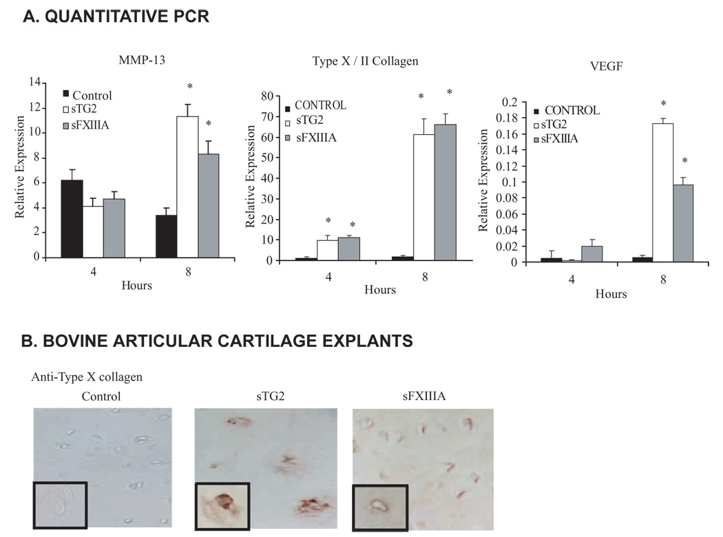
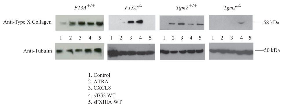
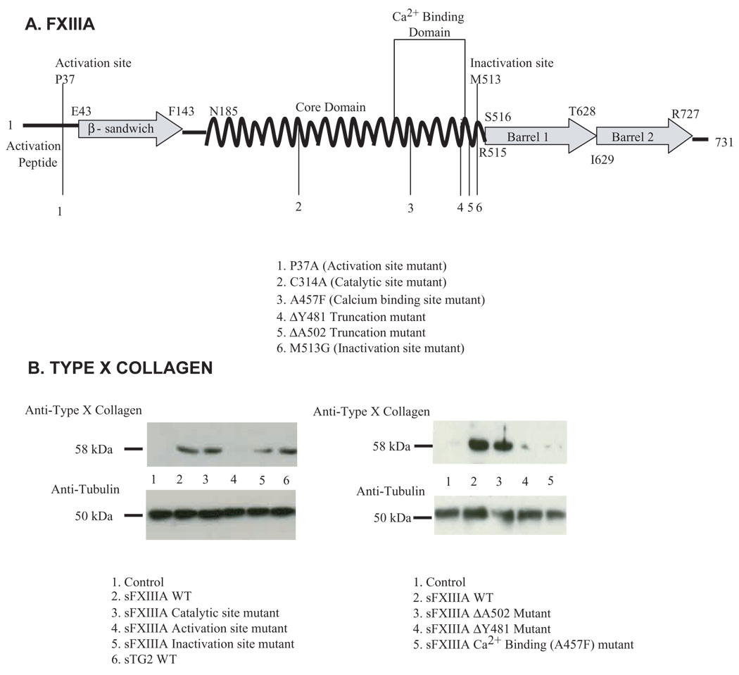
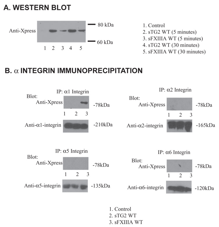
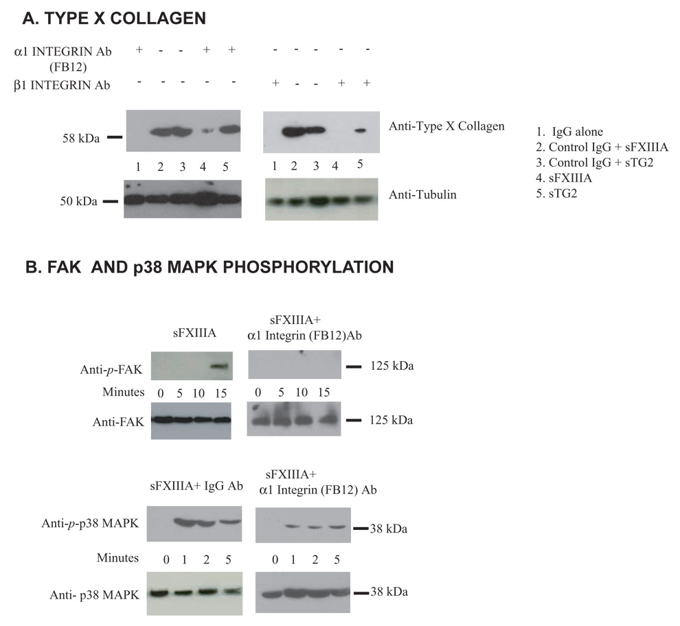
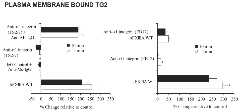
Two transglutaminases (TGs), factor XIIIA (FXIIIA) and TG2, undergo physiologic upregulation in growth plate hypertrophic chondrocytes, and pathological upregulation in osteoarthritic cartilage. Externalization of guanine-nucleotide-bound TG2 drives chondrocyte maturation to hypertrophy, a state linked to matrix remodeling and calcification. Here, we tested the hypothesis that FXIIIA also promotes hypertrophic differentiation. Using human articular chondrocytes, we determined that extracellular FXIIIA induced chondrocyte hypertrophy associated with rapid movement of TG2 to the cell surface. Site-directed mutagenesis revealed that FXIIIA Pro37 bordering the thrombin endoproteolytic Arg38-Gly39 site, but not intrinsic TG catalytic activity, were necessary for FXIIIA to induce chondrocyte hypertrophy. TGs have been demonstrated to interact with certain integrins and, during osteoarthritis (OA), alpha1beta1 integrin is upregulated and associated with hypertrophic chondrocytes. FXIIIA engaged alpha1beta1 integrin in chondrocytes. Antibody crosslinking of alpha1beta1 integrin mobilized TG2. Conversely, an alpha1beta1-integrin-specific blocking antibody inhibited the capacity of FXIIIA to induce TG2 mobilization to the cell surface, phosphorylation of p38 MAP kinase, and chondrocyte hypertrophy. Our results identify a unique functional network between two cartilage TG isoenzymes that accelerates chondrocyte maturation without requirement for TG-catalyzed transamidation by either TG.


Figures

Fig 1
Fig 1
Both exogenous FXIIIA and TG2 induce chondrocyte hypertrophic differentiation. We studied induction by TG2 and FXIIIA of MMP-13, VEGF, type X collagen and Syndecan-3 in cultured chondrocytes. Normal human knee articular chondrocytes plated in 12 well dishes (at 1 × 10⁵ cells/well) were incubated 4 or 8 hours with 100 ng/ml of recombinant wild type TG2 or FXIIIA in the ascorbate-containing medium A described in the Methods. (A) Using quantitative RT-PCR, we determined chondrocyte mRNA expression levels relative to GAPDH for MMP-13 and VEGF, and the mRNA expression ratio of type X : type II collagen, studying data collected from three separate human donors (n=9, *p<0.05). (B) Assessment of the induction by TG2 and FXIIIA of Type X collagen in cartilage organ culture. Normal bovine articular cartilage explants in organ culture were incubated with 100 ng/ml recombinant wild type TG2 or FXIIIA for 5 days in medium A, and 10 µm frozen sections were stained for type X collagen expression (representative of 5 donors).
Fig 2
Fig 2
FXIIIA-stimulated type X collagen expression is dependent upon TG2. Assessment of TG2 and FXIIIA knockout mouse cells. Primary knee chondrocytes were removed from F13A+/+, F13A−/− Tgm2+/+, and Tgm2−/− mice. After two weeks in culture, aliquots of 5×10³ cells in Medium A were stimulated for 5 days with 10 nM ATRA, 10 ng/ml CXCL8, or 100 ng/ml of sTG2 or sFXIIIA, and then type X collagen was examined by SDS-PAGE/Western blotting, as described in the Methods. Representative of three experiments.
Fig 3
Fig 3
(A) FXIIIA site directed mutants generated for structural analysis examination. The schematic depicts features of the primary structure of wild type (WT) FXIIIA and the panel of FXIIIA site directed mutants generated and studied. (B) Aliquots of human articular chondrocytes (1 × 10⁵ cells) were incubated with 100 ng/ml of each recombinant protein in medium A for 72 hours, and type X collagen assessed in cell lysates by SDS-PAGE/ Western blotting. Representative of three donors in three separate experiments (n=9).
Fig 4
Fig 4
Rapid mobilization of TG2 by Ca2+-bound FXIIIA is essential for stimulation of p38 phosphorylation. (A) Aliquots of 5 × 10³ human articular chondrocytes were starved in serum-free DMEM high glucose medium for 2 hours and then stimulated with WT or mutant sFXIIIA. TG2-specific antibodies were used to detect membrane bound TG2, quantified through successive incubations with biotin anti-rabbit and strepavidin-AP antibodies as described in the Methods. (n=9) (B) Aliquots of 3 × 10⁵ human articular chondrocytes were starved in serum free DMEM high glucose medium for 2 hours and then stimulated with WT or mutant sFXIIIA for the time indicated. Cell lysates were analyzed by Western blotting for p-p38 and total p38. Representative of three donors in three separate experiments (n=9).
Fig 5
Fig 5
Interaction of recombinant FXIIIA with human chondrocytes through α1 integrin subunit. (A) Aliquots of 1 × 10⁵ human articular chondrocytes were starved for 2 hours in serum free DMEM and then incubated for the indicated times with 100 ng/ml of sTG2 or sFXIIIA. Washed cell lysates were examined for the presence of the Xpress epitope on the recombinant proteins by SDS-PAGE/Western blotting. (B) Aliquots of 1 × 10⁶ human articular chondrocytes were incubated for three days in medium A containing 100 ng/ml of sTG2 or sFXIIIA where indicated. Cell lysates (200 µg protein) were immunoprecipitated using 1 µg/ml of α1 (clone TS2/7), α2, α5, or α6 integrin subunit-specific antibody, and precipitated proteins were analyzed for the Xpress tag or each respective α integrin subunit by Western blotting. Representative of 3 separate experiments using 3 different donors.
Fig 6
Fig 6
FXIIIA induction of type X collagen is associated with FAK and p38 MAP kinase phosphorylation and dependent upon interaction with the α1 integrin subunit. (A) Aliquots of 1 × 10⁵ human articular chondrocytes were starved and pretreated for 2 hours with 1 µg/ml of IgG control, β1 integrin subunit or α1 integrin subunit blocking (FB12) antibodies in serum free DMEM and then incubated for the indicated times with 100 ng/ml of sTG2 or sFXIIIA, with type X collagen assessed by Western blotting of cell lysates as above. (B) Aliquots of 1 × 10⁵ cells were starved for 2 hours in serum-free DMEM and then incubated for the indicated times with 100 ng/ml of sTG2 or sFXIIIA and 1 µg/ml of α1 integrin subunit-blocking antibody (FB12) where indicated, with cell lysates examined for FAK and p38 phosphorylation by Western blotting. Representative of results from 3 experiments employing 3 separate donors.
Fig 7
Fig 7
Rapid mobilization of TG2 to the cell surface by sFXIIIA and antibody crosslinking of α1 integrin subunit. To determine if FXIIIA-induced movement of TG2 to the cell surface is integrin dependent, aliquots of 5 × 10³ human articular chondrocytes were starved in serum-free DMEM high glucose medium for 2 hours and then stimulated with sFXIIIA, the α1 integrin subunit antibody, TS2/7 (with and without crosslinking by anti-mouse IgG) versus an IgG control antibody. Additionally, after starvation the chondrocytes were pretreated with the blocking α1 integrin subunit antibody, FB12 and then stimulated for 5 and 10 minutes with WT sFXIIIA. After fixation of the cells, TG2-specific antibodies were used to detect membrane bound TG2, quantified through successive incubations with biotin anti-rabbit and strepavidin-AP antibodies as described in the Methods.

References

    1. AbdAlla S, Lother H, Langer A, el Faramawy Y, Quitterer U. Factor XIIIA transglutaminase crosslinks AT1 receptor dimers of monocytes at the onset of atherosclerosis. Cell. 2004;119:343–354. - PubMed
    1. Aeschlimann D, Wetterwald A, Fleisch H, Paulsson M. Expression of tissue transglutaminase in skeletal tissues correlates with events of terminal differentiation of chondrocytes. J Cell Biol. 1993;20:1461–1470. - PMC - PubMed
    1. Aeschlimann D, Mosher D, Paulsson M. Tissue transglutaminase and factor XIII in cartilage and bone remodeling. Semin Thromb Hemost. 1996;22:437–443. - PubMed
    1. Akimov SS, Belkin AM. Cell-surface transglutaminase promotes fibronectin assembly via interaction with the gelatin-binding domain of fibronectin: a role in TGFbeta-dependent matrix deposition. J Cell Sci. 2001;114:2989–3000. - PubMed
    1. Akimov SS, Krylov D, Fleischman LF, Belkin AM. Tissue transglutaminase is an integrin-binding adhesion coreceptor for fibronectin. J Cell Biol. 2000;148:825–838. - PMC - PubMed

Publication types

MeSH terms