. 2008 Jun 17;105(24):8292-7.
doi: 10.1073/pnas.0707761105. Epub 2008 Jun 10.

The p110beta isoform of phosphoinositide 3-kinase signals downstream of G protein-coupled receptors and is functionally redundant with p110gamma

Affiliations

The p110beta isoform of phosphoinositide 3-kinase signals downstream of G protein-coupled receptors and is functionally redundant with p110gamma

Julie Guillermet-Guibert et al. Proc Natl Acad Sci U S A. .

Abstract

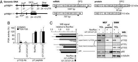
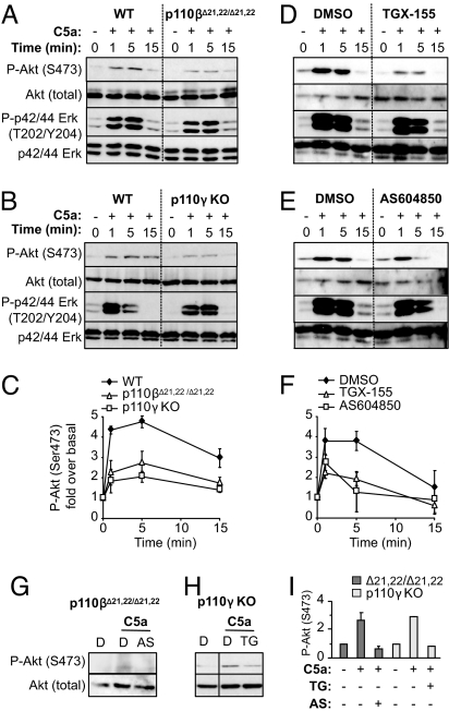
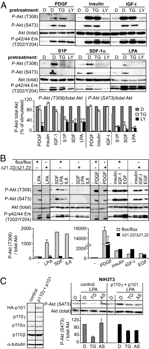
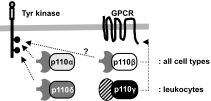
The p110 isoforms of phosphoinositide 3-kinase (PI3K) are acutely regulated by extracellular stimuli. The class IA PI3K catalytic subunits (p110alpha, p110beta, and p110delta) occur in complex with a Src homology 2 (SH2) domain-containing p85 regulatory subunit, which has been shown to link p110alpha and p110delta to Tyr kinase signaling pathways. The p84/p101 regulatory subunits of the p110gamma class IB PI3K lack SH2 domains and instead couple p110gamma to G protein-coupled receptors (GPCRs). Here, we show, using small-molecule inhibitors with selectivity for p110beta and cells derived from a p110beta-deficient mouse line, that p110beta is not a major effector of Tyr kinase signaling but couples to GPCRs. In macrophages, both p110beta and p110gamma contributed to Akt activation induced by the GPCR agonist complement 5a, but not by the Tyr kinase ligand colony-stimulating factor-1. In fibroblasts, which express p110beta but not p110gamma, p110beta mediated Akt activation by the GPCR ligands stromal cell-derived factor, sphingosine-1-phosphate, and lysophosphatidic acid but not by the Tyr kinase ligands PDGF, insulin, and insulin-like growth factor 1. Introduction of p110gamma in these cells reduced the contribution of p110beta to GPCR signaling. Taken together, these data show that p110beta and p110gamma can couple redundantly to the same GPCR agonists. p110beta, which shows a much broader tissue distribution than the leukocyte-restricted p110gamma, could thus provide a conduit for GPCR-linked PI3K signaling in the many cell types where p110gamma expression is low or absent.


Conflict of interest statement

Conflict of interest: B.V. is a consultant for PIramed (Slough, UK) and AstraZeneca.

Figures

Fig. 1.
Fig. 1.
Genetic inactivation of p110β in mice. (A) Schematic representation of genomic DNA, mRNA, and protein primary structure of the p110βflox and p110βΔ21,22 locus. Exons 22 and 23 of Pik3cb were flanked by loxP sites as described in Fig. S1, creating a floxed p110β allele that is further referred to as p110βflox. Exon 22 encodes the DFG domain and activation loop that are critical for p110β kinase activity. Constitutive deletion of exons 21 and 22 in mice was achieved by intercrossing p110βflox/flox mice with Cre deleter mice, creating the p110βΔ21,22 allele. The loxP sites were positioned in such a way that, after treatment with Cre, the locus is expected to give rise to mRNA in which exon 20 is spliced in-frame onto exon 23, now encoding an internally truncated p110β protein lacking amino acids 886–975 in the catalytic domain. This p110βΔ21,22 protein is predicted to be kinase-inactive, with a ≈10-kDa reduction in Mr relative to the WT p110β protein and to retain reactivity with antisera raised against the extreme C terminus of p110β. (Left) Exon sequences are represented by filled black rectangles, and intron sequences are indicated by a horizontal black line. The loxP sites are shown as hatched triangles with the pointed end indicating orientation. (Center) Exon boundaries are represented from exons 16 to 24. The positions of the primers used for PCR screening are designated by arrows together with the expected amplification products and their size (bp). (Right) Schematic representation and predicted Mr of WT and mutant p110β proteins, based on exon boundaries from exons 16 to 24. (B) Effect of p110β deletion on in vitro lipid kinase activity. Homogenates of the indicated MEFs were immunoprecipitated by using p110β Abs or absorbed onto PDGF receptor phosphoTyr peptide (pY peptide) immobilized to Sepharose (which binds all class IA PI3K regulatory subunits), followed by in vitro lipid kinase assay with or without 100 nM TGX-155. The level of p110β and p85 in the indicated cell fractions was verified by immunoblotting (data not shown). (C) Effect of p110β deletion on PI3K isoform expression. (Left) Analysis of p110β protein expression in MEFs from p110βflox/flox, p110βflox/Δ21,22, and p110βΔ21,22/Δ21,22 mice by Western blotting. Immunoblot signal intensities obtained with Abs to p110α, p110β, and p85 were expressed relative to signals obtained with antibodies to α-tubulin for at least three different embryos and expressed relative to the signals in p110βflox/flox MEFs. Quantification of the signals detected by the sc-602 p110β Abs revealed that only the intensity of the upper band is decreased upon Cre treatment, whereas the intensity of the lower band is unaffected. p110α expression was not affected in each genotype. p110γ and p110δ were hardly detectable in fibroblasts and could not be reliably quantified. (Right) Total cell lysates or pY-peptide pull-downs were immunoblotted by using the indicated Abs. In NIH 3T3 cells and MEFs (but not in BMMs), the anti-p110β Abs used for Western blotting (sc-602) recognize a nonspecific protein just below the specific p110β signal, indicated as lower band (aspecific) and upper band (p110β), respectively. It is only the upper band that disappears upon p110β deletion.
Fig. 2.
Fig. 2.
Role of p110β and p110γ in cell signaling in macrophages. (A and B) Starved BMMs of the indicated genotype were stimulated with C5a, followed by immunoblotting using the indicated Abs. (D and E) Starved WT BMMs were treated for 30 min with TGX-155 (0.5 μM) (D) or AS604850 (1 μM) (E), followed by C5a stimulation and immunoblotting using the indicated Abs. (C and F) Quantification of at least two independent experiments was performed, and data are shown as fold over P-Akt under unstimulated conditions. (G–I) Starved BMMs of the indicated genotype were treated for 30 min with AS604850 (1 μM) or TGX-155 (0.5 μM) and stimulated for 5 min with C5a, followed by immunoblotting using the indicated Abs. I shows the quantification of the experiments in G and H. Data are expressed as fold over P-Akt under unstimulated conditions.
Fig. 3.
Fig. 3.
Role of p110β and p110γ in cell signaling in fibroblasts. (A) (Top and Middle) Starved NIH 3T3 cells were treated for 1 h with TGX-155 (TG; 1 μM), LY294002 (LY; 20 or 5 μM), or vehicle DMSO (D) followed by stimulation for 10 min with the indicated ligands. Total cell lysates were immunoblotted with the indicated Abs. A representative immunoblot is shown. (Bottom) Quantification of three independent experiments was performed, and data are presented as percentage of ligand-induced P-Akt/total Akt. (B) Starved MEFs of indicated genotype were stimulated with indicated stimuli for 10 min and immunoblotted with the indicated Abs. A representative experiment performed with MEFs isolated from different embryos is shown. (C) p110 isoform expression in NIH 3T3 cells transfected with empty vector (NIH 3T3 control) or p110γ and its regulatory subunit HA-p101 (p110γ + p101). Starved cells were treated for 1 h with TGX-155 (1 μM) or AS604850 (1 μM), followed by stimulation for 10 min with LPA and immunoblotting with the indicated Abs.
Fig. 4.
Fig. 4.
Model for class IA PI3K signaling downstream of Tyr kinase receptors and GPCRs. The ubiquitously expressed p110α plays an important role in Tyr kinase-driven PI3K signaling in all cell types, with p110δ providing additional Tyr kinase-driven PI3K signaling in cell types in which it is expressed at a high level, such as in leukocytes. A similar scenario could be envisaged for the broadly expressed p110β that could control GPCR-driven PI3K in all cell types, with p110γ providing additional GPCR-PI3K signaling capacity in white blood cells.

References

    1. Vanhaesebroeck B, Leevers SJ, Panayotou G, Waterfield MD. Phosphoinositide 3-kinases: A conserved family of signal transducers. Trends Biochem Sci. 1997;22:267–272. - PubMed
    1. Bi L, Okabe I, Bernard DJ, Nussbaum RL. Early embryonic lethality in mice deficient in the p110β catalytic subunit of PI 3-kinase. Mamm Genome. 2002;13:169–172. - PubMed
    1. Jackson SP, et al. PI 3-kinase p110β: A new target for antithrombotic therapy. Nat Med. 2005;11:507–514. - PubMed
    1. Kurosu H, et al. Heterodimeric phosphoinositide 3-kinase consisting of p85 and p110β is synergistically activated by the β-γ subunits of G proteins and phosphotyrosyl peptide. J Biol Chem. 1997;272:24252–24256. - PubMed
    1. Maier U, Babich A, Nurnberg B. Roles of noncatalytic subunits in gbetagamma-induced activation of class I phosphoinositide 3-kinase isoforms β and γ. J Biol Chem. 1999;274:29311–29317. - PubMed

Publication types

MeSH terms

Substances