

Paratracheal air cysts: a common finding on routine CT examinations of the cervical spine and neck that may mimic pneumomediastinum in patients with traumatic injuries
- PMID: 18544671
- PMCID: PMC8118841
- DOI: 10.3174/ajnr.A1058
Paratracheal air cysts: a common finding on routine CT examinations of the cervical spine and neck that may mimic pneumomediastinum in patients with traumatic injuries
Abstract
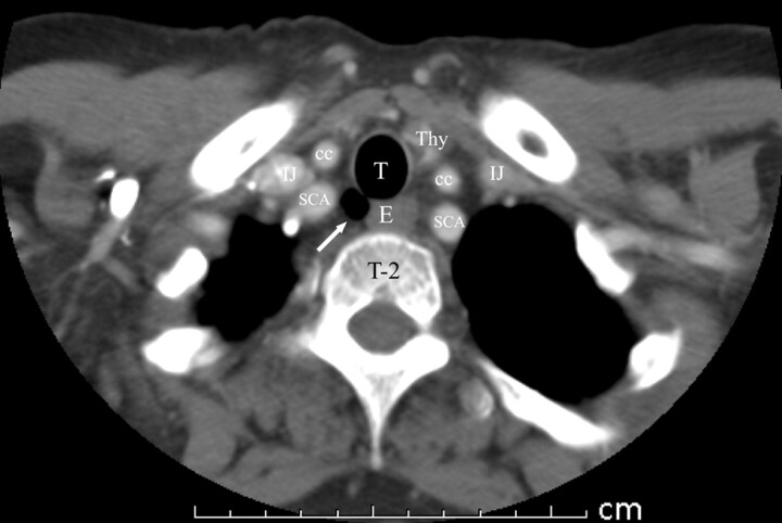
Background and purpose: Collections of extraluminal paratracheal gas may be present on CT images of the neck and cervical spine and the radiologist may question whether this is related to a pathologic process. This study is designed to demonstrate the appearance, clinical presentation, and prevalence of paratracheal air cysts, which, on CT examinations of the neck, can mimic abnormal extraluminal air.
Materials and methods: From January 1, 2005, through May 22, 2005, a total of 702 CT examinations of the cervical spine or soft tissue of the neck were reviewed. All examinations were at 2- to 5-mm thickness. Sagittal and coronal reconstructions were available for review, if necessary. Paratracheal air cysts were evaluated for size; the presence of visible communication with the trachea; association with pneumothorax, pneumomediastinum, or subcutaneous emphysema; and association with findings of emphysematous changes in the lung apices. Patient demographics of age, sex, and whether the patient had sustained a traumatic injury were also collected.
Results: Of the 702 patients evaluated, 26 (3.7%) had paratracheal air cysts, all of which were found on the right, at the level of the thoracic inlet. Ages of the patients ranged from 15 to 74 years. In 9 (34.6%) of the patients, a direct communication with the trachea was seen. Sizes of the paratracheal air cysts ranged from 1 x 2 mm to 10 x 15 mm. No association was found with CT findings of emphysema in the lung apices, abnormal soft tissue air, or trauma.
Conclusion: Right paratracheal air cysts are a common CT finding that occur in a predictable location. In the setting of trauma, these characteristic structures can mimic pneumomediastinum and are seen in approximately 3% to 4% of the US population. The cause is unclear but may be either congenital or an acquired phenomenon, given that they are often seen in both children and adults. We found no association with either trauma or the presence of emphysematous changes in the lung apices.
Figures


References
-
- Goo JM, Im JG, Ahn JM, et al. Right paratracheal air cysts in the thoracic inlet: clinical and radiologic significance. AJR Am J Roentgenol 1999;173:65–70 - PubMed
-
- Early EK, Bothwell MR. Congenital tracheal diverticulum. Otolaryngol Head Neck Surg 2002;127:119–21 - PubMed
-
- Restrepo S, Villamil MA, Rojas IC, et al. Association of two respiratory congenital anomalies: tracheal diverticulum and cystic adenomatoid malformation of the lung. Pediatr Radiol 2004;34:263–66 - PubMed
-
- Infante M, Mattavelli F, Valente M, et al. Tracheal diverticulum: a rare cause and consequence of chronic cough. Eur J Surg 1994;160:315–16 - PubMed
-
- Tanaka H, Mori Y, Kurokawa K, et al. Paratracheal air cysts communicating with the trachea: CT findings. J Thoracic Imag 1997;12:38–40 - PubMed
MeSH terms
LinkOut - more resources
Full Text Sources
Medical