. 2008 Jun 11;3(6):e2451.
doi: 10.1371/journal.pone.0002451.

Inducible ablation of melanopsin-expressing retinal ganglion cells reveals their central role in non-image forming visual responses

Affiliations

Inducible ablation of melanopsin-expressing retinal ganglion cells reveals their central role in non-image forming visual responses

Megumi Hatori et al. PLoS One. .

Erratum in

  • PLoS ONE. 2008 Jun;4(6). doi: 10.1371/annotation/c02106ba-b00b-4416-9834-cf0f3ba49a37. Buch, Thorsten [added]; Waisman, Ari [added]
  • PLoS ONE. 2008;3(7). doi: 10.1371/annotation/16f913dd-c33b-419f-9555-c788c80c189f

Abstract

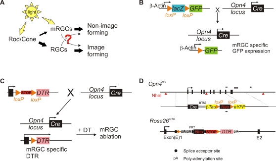
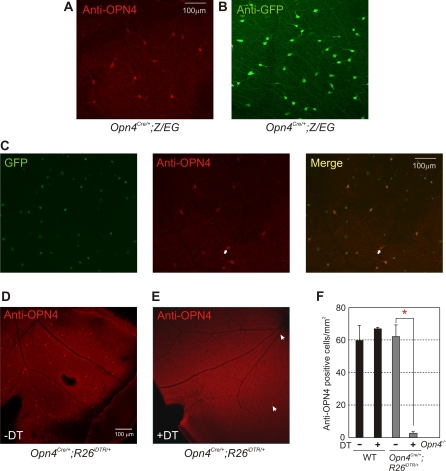
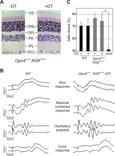
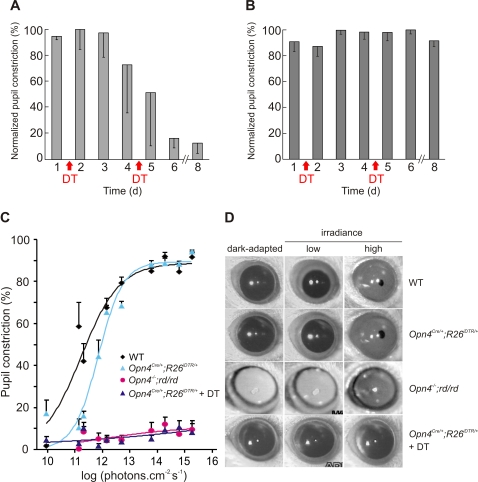
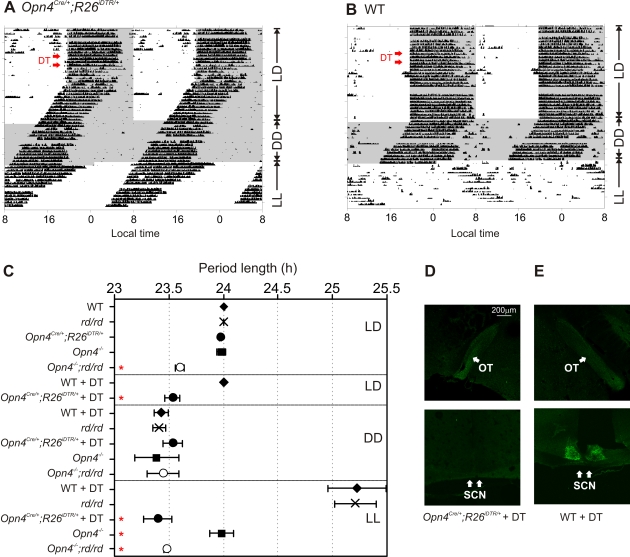
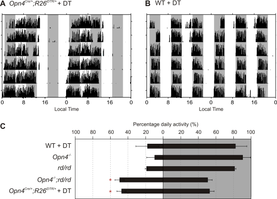
Rod/cone photoreceptors of the outer retina and the melanopsin-expressing retinal ganglion cells (mRGCs) of the inner retina mediate non-image forming visual responses including entrainment of the circadian clock to the ambient light, the pupillary light reflex (PLR), and light modulation of activity. Targeted deletion of the melanopsin gene attenuates these adaptive responses with no apparent change in the development and morphology of the mRGCs. Comprehensive identification of mRGCs and knowledge of their specific roles in image-forming and non-image forming photoresponses are currently lacking. We used a Cre-dependent GFP expression strategy in mice to genetically label the mRGCs. This revealed that only a subset of mRGCs express enough immunocytochemically detectable levels of melanopsin. We also used a Cre-inducible diphtheria toxin receptor (iDTR) expression approach to express the DTR in mRGCs. mRGCs develop normally, but can be acutely ablated upon diphtheria toxin administration. The mRGC-ablated mice exhibited normal outer retinal function. However, they completely lacked non-image forming visual responses such as circadian photoentrainment, light modulation of activity, and PLR. These results point to the mRGCs as the site of functional integration of the rod/cone and melanopsin phototransduction pathways and as the primary anatomical site for the divergence of image-forming and non-image forming photoresponses in mammals.


Conflict of interest statement

Competing Interests: The authors have declared that no competing interests exist.

Figures

Figure 1
Figure 1. Strategy for fluorescent labeling or inducible ablation of mRGC lineage in the mouse retina.
(A) The cellular circuitry underlying the rod/cone and mRGC contribution to visual responses. Thickness of the filled arrows roughly highlights the relative strength of information flow. (B) Strategy to fluorescently label mRGCs by breeding a mouse carrying a Cre recombinase “knocked-in” to the melanopsin promoter to Z/EG mouse that allows Cre-dependent expression of GFP from chicken beta-actin promoter. (C) Strategy to achieve inducible and specific ablation of mRGCs. Opn4Cre/+ mouse was bred to a mouse expressing Cre-dependent expression of simian diphtheria toxin receptor. The resulting progeny develop normal mRGCs expressing DTR, which allows specific ablation of these cells by DT administration. Schematic of two targeting vectors used to achieve inducible mRGC ablation are shown in (D). The Cre knock-in cassette for targeted insertion to melanopsin locus also carried coding sequences for CRE dependent expression of βTau-eYFP. However, fluorescence from βTau:eYFP was undetectable in retina from Opn4Cre/+ mice (data not shown). The targeting vector and generation of R26iDTR/+ mice are described in . A schematic of the targeting vector is shown here.
Figure 2
Figure 2. Cre-dependent GFP labeling and inducible ablation of mRGCs.
Retina of adult Opn4Cre/+;Z/EG mouse probed with (A) anti-OPN4 or (B) anti-GFP antibodies show staining of a small subset of cells. (C) Only a subset of GFP positive cells also stained with anti-OPN4 antibody. A section of the flat mount retina containing a cell (marked with an arrow) that stained with melanopsin antibody, but did not express detectable level of GFP is shown. (D) Retina of Opn4Cre/+;R26iDTR/+ showed normal melanopsin immunostaining in a small fraction of RGCs. (E) Two weeks after DT administration, the number of melanopsin-immunoreactive cells were significantly reduced. (F) Average melanopsin immunoreactive cell density in WT and Opn4Cre/+;R26iDTR/+ retina was comparable. Two weeks after DT administration, the number of cells in WT retina remained unchanged, while that in Opn4Cre/+;R26iDTR/+ retina reduced from 62.3 mm−2 to 2.8 mm−2. No melanopsin immunostaining was observed in Opn4−/− mice. It is important to note that retina from all DT-treated mice tested by immunostaining still retained a few melanopsin staining cells (arrows), implying incomplete expression of Cre in all mRGCs and/or insufficient level of bioavailable DT. Average cell counts (+SEM, n = 3 to 5 retinas) from each genotype/treatment group are shown. Significant difference in cell numbers (Student's t test, p<0.05) between mRGCs without and with DT was highlighted with an asterisk.
Figure 3
Figure 3. mRGC ablation does not alter the normal retina architecture and image-forming responses.
(A) Hematoxylin and Eosin staining of 5 μm thick paraffin embedded sections of retina from Opn4Cre/+;R26iDTR/+ mice without and with DT injection. DT application had no detectable adverse effect on the normal stratification of the retina (outer segment (OS), outer nuclear layer (ONL), outer plexiform layer (OPL), inner nuclear layer (INL), inner plexiform layer (IPL), and ganglion cell layer (GCL)). (B) Representative full-field ERG of WT and DT-treated Opn4Cre/+;R26iDTR/+ mice showing rod, cone and maximal combined responses. Responses from both eyes were simultaneously measured and plotted. Quantitative analysis of magnitude and timing of a-wave, b-wave and oscillatory potentials of these two genotype groups (3 mice each) showed no significant difference (data not shown). (C) Image forming visual function as assessed by the visual cliff test was unaffected by mRGC ablation. Average percentage (+SEM, n = 5 to 13 mice) of positive choice in 10 trials for each mouse are shown. Mice with outer retina degeneration (rd/rd) made random choices while stepping down from the platform and were significantly different (Student's t test, p<0.05; red asterisk) from the other four groups. No significant difference in test performance was found among native or DT-treated WT or Opn4Cre/+;R26iDTR/+ mice.
Figure 4
Figure 4. Necessity of mRGCs for PLR.
DT injection severely attenuates pupil constriction in response to 20 μW of monochromatic blue light (470 nm) in Opn4Cre/+;R26iDTR/+ mice (A), but does not affect in such PLR response in WT mice (B). Normalized pupil constriction (-SD; n = 3 mice) measured one day prior to or every day following DT injection for up to 8 days are shown. There was variability in the rate of loss in PLR response among Opn4Cre/+;R26iDTR/+ as reflected in the larger error bars. (C) Pupil constriction in response to varying irradiance levels over 5 log units shows the necessity of mRGC for PLR. Average (+SEM, n = 5 to 6 mice) and fitted sigmoid curves for WT, Opn4Cre/+;R26iDTR/+, Opn4−/−;rd/rd and DT-treated Opn4Cre/+;R26iDTR/+ mice are shown. (D) Representative frozen video images showing dark adapted pupil and pupil under low (1011 photons.cm−2.s−1) or high intensity light (1015 photons.cm−2.s−1) of 470 nm are shown. Notice the complete lack of pupil constriction in Opn4−/−;rd/rd and in DT-treated Opn4Cre/+;R26iDTR/+ mice. For each genotype representative images of the same eye under three different conditions are shown.
Figure 5
Figure 5. mRGCs are necessary for light adaptation of circadian wheel running activity rhythm.
Representative daily wheel running activity profile of an (A) Opn4Cre/+;R26iDTR/+ and (B) WT mouse under 12 h light∶12 h dark (LD), constant dark (DD) or constant light (LL) are shown. A week after DT injection (red arrows) the wheel running activity of Opn4Cre/+;R26iDTR/+ began to “free run” with a periodicity similar to that under no light. Constant light had no effect on the daily drift in activity onset in this mouse. The wheel running activity of the WT mouse remained entrained to the LD cycle even after DT injection and was lengthened under constant light. Daily wheel running activity profile of mice were binned in 6 min and double plotted such that the activity from consecutive days are plotted to the right and beneath the data from previous day. Periods of darkness are shown in shaded area. (C) Genotypes of mice and their respective average (±SEM, n = 3 to 6 mice) period length of wheel running activity rhythm under conditions of LD, DD and LL as determined by periodogram analysis in Clocklab software are shown. Within each lighting or treatment group (separated by solid box) significant difference (Student's t test, p<0.05) from WT group is shown by red asterisk. (D) Anterograde CTB-Alexa Fluor 488 tracing in the optic tract (OT) is intact, but is completely abolished in the SCN of DT treated Opn4Cre/+;R26iDTR/+ mice. (E) Staining in both regions are left intact in WT mice treated with DT.
Figure 6
Figure 6. Lack of negative masking in mRGC ablated mice.
Representative wheel running activity profile of DT-treated (A) Opn4Cre/+;R26iDTR/+ and (B) WT mice held under ultradian cycle of 4 h light and 4 h darkness are shown. The sessions of darkness are shown by shaded area. Activity profile is plotted as in Figure 5A. (C) Average percentage (±SEM, n = 3 to 6 mice) of daily activity during the bouts of light sessions (on left) or during darkness (on right) for five different groups of mice are shown. Groups with percent activity during light phase significantly different (Student's t test, p<0.05) from that of the DT-treated WT mice are shown with asterisk.

References

    1. Provencio I, Rodriguez IR, Jiang G, Hayes WP, Moreira EF, et al. A novel human opsin in the inner retina. J Neurosci. 2000;20:600–605. - PMC - PubMed
    1. Gooley JJ, Lu J, Chou TC, Scammell TE, Saper CB. Melanopsin in cells of origin of the retinohypothalamic tract. Nat Neurosci. 2001;4:1165. - PubMed
    1. Berson DM, Dunn FA, Takao M. Phototransduction by retinal ganglion cells that set the circadian clock. Science. 2002;295:1070–1073. - PubMed
    1. Hattar S, Liao HW, Takao M, Berson DM, Yau KW. Melanopsin-containing retinal ganglion cells: architecture, projections, and intrinsic photosensitivity. Science. 2002;295:1065–1070. - PMC - PubMed
    1. Belenky MA, Smeraski CA, Provencio I, Sollars PJ, Pickard GE. Melanopsin retinal ganglion cells receive bipolar and amacrine cell synapses. J Comp Neurol. 2003;460:380–393. - PubMed

Publication types