. 2008 Jun 11;3(6):e2337.
doi: 10.1371/journal.pone.0002337.

Drug metabolism in human brain: high levels of cytochrome P4503A43 in brain and metabolism of anti-anxiety drug alprazolam to its active metabolite

Affiliations

Drug metabolism in human brain: high levels of cytochrome P4503A43 in brain and metabolism of anti-anxiety drug alprazolam to its active metabolite

Varsha Agarwal et al. PLoS One. .

Abstract

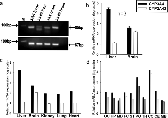
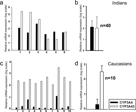
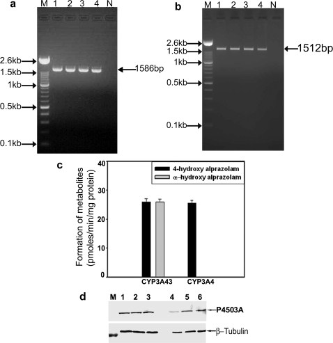
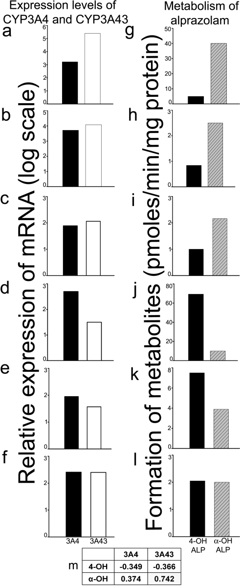
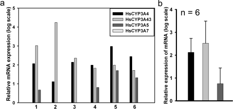
Cytochrome P450 (P450) is a super-family of drug metabolizing enzymes. P450 enzymes have dual function; they can metabolize drugs to pharmacologically inactive metabolites facilitating their excretion or biotransform them to pharmacologically active metabolites which may have longer half-life than the parent drug. The variable pharmacological response to psychoactive drugs typically seen in population groups is often not accountable by considering dissimilarities in hepatic metabolism. Metabolism in brain specific nuclei may play a role in pharmacological modulation of drugs acting on the CNS and help explain some of the diverse response to these drugs seen in patient population. P450 enzymes are also present in brain where drug metabolism can take place and modify therapeutic action of drugs at the site of action. We have earlier demonstrated an intrinsic difference in the biotransformation of alprazolam (ALP) in brain and liver, relatively more alpha-hydroxy alprazolam (alpha-OHALP) is formed in brain as compared to liver. In the present study we show that recombinant CYP3A43 metabolizes ALP to both alpha-OHALP and 4-hydroxy alprazolam (4-OHALP) while CYP3A4 metabolizes ALP predominantly to its inactive metabolite, 4-OHALP. The expression of CYP3A43 mRNA in human brain samples correlates with formation of relatively higher levels of alpha-OH ALP indicating that individuals who express higher levels of CYP3A43 in the brain would generate larger amounts of alpha-OHALP. Further, the expression of CYP3A43 was relatively higher in brain as compared to liver across different ethnic populations. Since CYP3A enzymes play a prominent role in the metabolism of drugs, the higher expression of CYP3A43 would generate metabolite profile of drugs differentially in human brain and thus impact the pharmacodynamics of psychoactive drugs at the site of action.


Conflict of interest statement

Competing Interests: The authors have declared that no competing interests exist.

Figures

Figure 1
Figure 1. Constitutive expression of CYP3A43 mRNA in human brain cortex.
(a) Total RNA was extracted from human brain cortex of 2 subjects and the 3′-UTR of CYP3A43 was amplified by RT-PCR (lanes 1 and 2). Lane N denoted reaction performed without template DNA. The size of the PCR product was 85bp. (b) Total RNA from human brain cortex was electrophoresed, transferred to positively charged nylon membrane and hybridized with antisense riboprobe prepared using the partial cDNA to CYP3A43. The mobility of 18S and 28S ribosomal RNA on the agarose gel is indicated.
Figure 2
Figure 2. Localization of CYP3A43 mRNA in human brain by fluorescence in situ hybridization.
(a) Intense fluorescence was seen in neuronal cell layers of frontal cortex. Differential localization was seen delineating the laminar architecture of the cortex. Bar = 100 µm (b) Higher magnification of cortical neurons (arrow). Bar = 25 µm (c) Control section hybridized with sense probe did not show any fluorescence. Bar = 100 µm (d) Reticular neurons in the midbrain expressed CYP3A43 mRNA. Bar = 100 µm (e) Fluorescent labelling of the granule cell layer (GL) in human cerebellum. Sparse staining was observed in the molecular layer (ML). Bar = 100 µm (f) Higher magnification of neurons of the granule cell layer in cerebellum. Bar = 25 µm (g) Control section hybridised with sense probe. Bar = 100 µm (h) CYP3A43 expression was seen in pyramidal neurons of CA1 in the hippocampus. Bar = 50 µm (i) Higher magnification of CA1 neurons. Bar = 25 µm (j) Robust staining of the CA2 pyramidal cell layer (arrow) of the hippocampus. Bar = 50 µm (k) Higher magnification of pyramidal neurons in the CA2 subfield. Bar = 25 µm (l) Intense fluorescence was seen in the CA3 pyramidal neurons (arrow head) of the human brain. Bar = 50 µm (m) Higher magnification of CA3 neurons (arrow head). Bar = 25 µm (n) Intense fluorescence was observed in the dentate gyrus. Staining of the interneurons of the hilus (arrow head) was also observed. Bar = 100 µm (o) Higher magnification of the granule cells in the dentate gyrus. Bar = 25 µm (p) Control section of hippocampus hybridized with sense probe did not show any fluorescence. Bar = 100 µm
Figure 3
Figure 3. Quantitative assessment of the expression of CYP3A4 and CYP3A43 in human brain cortex.
(a) RT-PCR amplification of the 85bp of 3′-UTR of CYP3A43 and 78bp of 3′-UTR of CYP3A4 using human brain and liver RNA. (b) qRT-PCR analysis using human liver and brain RNA (n = 3) showed the relative higher expression of CYP3A43 in brain compared to liver from the same individuals. Quantitation of the relative expression of CYP3A4 and CYP3A43 in (c) different human brain tissues (n = 1) and (d) from different regions of the brain from same individual (n = 3).
Figure 4
Figure 4. Expression of CYP3A43 and CYP3A4 in human brain samples from different population groups.
Expression of CYP3A43 and CYP3A4 in 6 individual samples of human brain cortex obtained from Indians depict the inter-individual variation (a). Expression of CYP3A43 and CYP3A4 averaged across 40 samples of human brain cortex from India (b). Comparison of CYP3A4 and CYP3A43 transcripts among 10 Caucasian brain samples (c) demonstrated that on average (d) CYP3A43 expression is 170 fold higher than CYP3A4. All samples were normalized using 18S rRNA.
Figure 5
Figure 5. Cloning and expression CYP3A43 and CYP3A4 and metabolism of alprazolam, in vitro by recombinant enzymes.
The complete open reading frame of CYP3A43 and CYP3A4 were amplified by RT-PCR using total RNA from four autopsy human brain samples. The amplicons of 1586bp and 1512bp were generated for CYP3A4 and CYP3A43, respectively (a, and b, from lanes 1–4). M represents 100bp ladder and Ne, the control reaction performed without template DNA. The amplicons were cloned into pcDNA 3.1 for expression in COS-1 cells. (c) Recombinant CYP3A43 metabolized alprazolam to α-OHALP and 4-OHALP in similar amounts (26 pmoles/min/mg protein), while recombinant CYP3A4 metabolized alprazolam only to 4-OHALP. The values are mean ISD (n = 3 independent experiments). (d) Immunoblot of recombinant CYP3A43 and CYP3A4 normalized using β-tubulin.
Figure 6
Figure 6. Metabolism of alprazolam to α-OHALP and 4-OHALP by human brain, in vitro.
The expression levels of CYP3A4 and CYP3A43 were compared with the rates of formation of α-OHALP and 4-OHALP in samples of cortex obtained from 6 human brain subjects. Subjects represented in (a, b, c) expressed high levels of CYP3A43 and relatively higher amounts of α-OHALP was formed compared to 4-OHALP (g, h, i). Subjects (d, e) expressed high levels of CYP3A4 and formed relatively higher amounts of 4-OHALP (j, k). Subject (f) showed similar levels of expression CYP3A4 and CYP3A43 which was reflected in the amount of hydroxylated metabolite formed. Pearson correlation analysis showed high degree of correlation between the expression levels of CYP3A43 and amounts of α-OHALP formed (m; r = 0.74).
Figure 7
Figure 7. Relative expression of functional genes of CYP3A subfamily members in human brain samples.
(a) The levels of CYP3A4, CYP3A5, CYP3A7, and CYP3A43 mRNA were quantified by qRT-PCR in the 6 human brain subjects. The expression of CYP3A5 was significantly lower then that of CYP3A4 and CYP3A43 and was detected only in four out of six samples. CYP3A7 was not detected in any of the subjects. (b) The average of the six samples analyzed.
Figure 8
Figure 8. Amplification of exonic regions of the ORF of CYP3A43 and CYP3A4 mRNA by RT-PCR.
(a) The schematic representation of 13 exons of CYP3A43 along with their splice variants detected by RT-PCR is depicted. (b) A 432bp amplicon was amplified spanning the exons 1–5 (lanes 1 and 2). (c) Two amplicons (594bp and 450bp) were generated when exons 6–10 were amplified (lanes 1 and 2). The 594bp product was the expected amplicon while the other amplicon (450bp) was a mixture of two PCR products of which one had a deletion of exon 7 and other had a deletion of exon 8. (d) Amplification of the region spanning exons 11–13 gave two amplicons of sizes 486bp and 350bp. The 486bp product was the expected size while 350bp product represented a splice variant that had the deletion of exon 12. (e) Amplification of exons 6–13 generated an amplicon (950bp) representing a splice variant having deletion of both exon 7 and 8 and partial inclusion of 121bp intron 7.

References

    1. Meyer RP, Gehlhaus M, Knoth R, Volk B. Expression and function of cytochrome P450 in brain drug metabolism. Curr Drug Metab. 2007;4:297–306. - PubMed
    1. Haining RL, Nichols HM. Cytochrome P450-catalyzed pathways in human brain: metabolism meets pharmacology or old drugs with new mechanism of action? Pharmacol Ther. 2007;3:537–545. - PubMed
    1. Anandatheerthavarada HK, Shankar SK, Ravindranath V. Rat brain cytochromes P-450: catalytic, immunochemical properties and inducibility of multiple forms. Brain Res. 1990;536:339–343. - PubMed
    1. Pai HV, Upadhya SC, Chinta SJ, Hedge SN, Ravindranath V. Differential metabolism of alprazolam by liver and brain cytochrome (P4503A) to pharmacologically active metabolite. Pharmacogenomics J. 2002;2:243–258. - PubMed
    1. Ravindranath V, Anandatheerthavarada HK, Shankar SK. Xenobiotic metabolism in human brain—presence of cytochrome P-450 and associated mono-oxygenases in human brain regions. Brain Res. 1989;496:331–335. - PubMed

Publication types

MeSH terms