. 2008 Jul;38(7):1814-21.
doi: 10.1002/eji.200838346.

TGF-beta-induced Foxp3+ regulatory T cells rescue scurfy mice

Affiliations

TGF-beta-induced Foxp3+ regulatory T cells rescue scurfy mice

Eva N Huter et al. Eur J Immunol. 2008 Jul.

Abstract

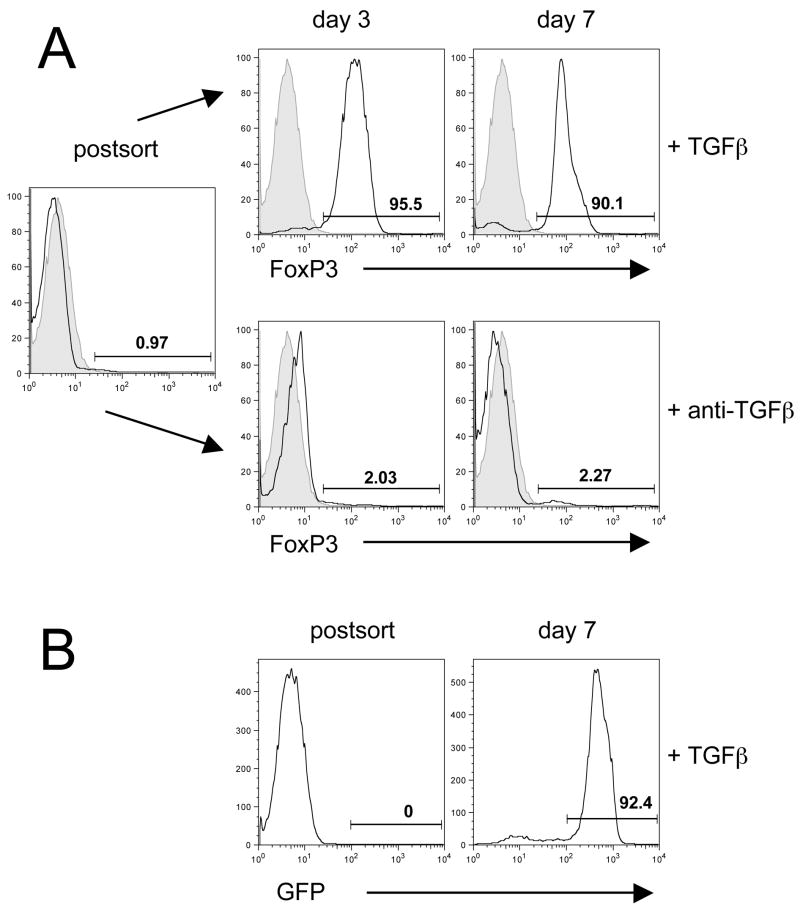
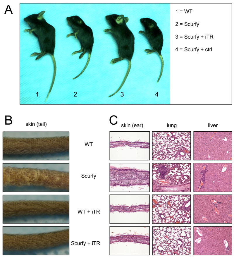
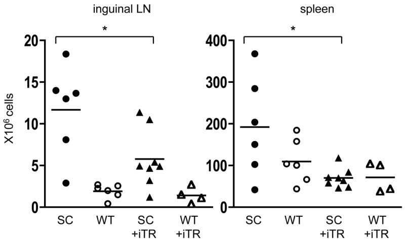
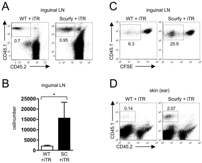
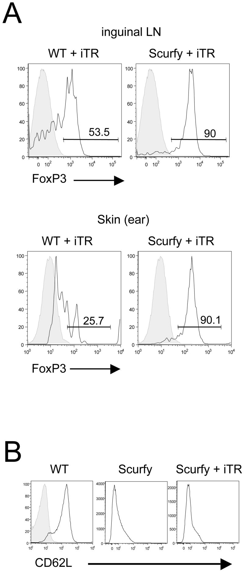
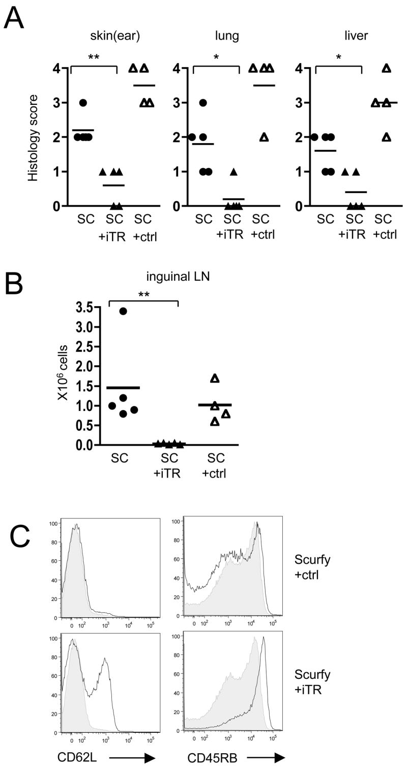
Scurfy mice have a deletion in the forkhead domain of the forkhead transcription factor p3 (Foxp3), fail to develop thymic-derived, naturally occurring Foxp3+ regulatory T cells (nTreg), and develop a fatal lymphoproliferative syndrome with multi-organ inflammation. Transfer of thymic-derived Foxp3+ nTreg into neonatal Scurfy mice prevents the development of disease. Stimulation of conventional CD4+Foxp3(-) via the TCR in the presence of TGF-beta and IL-2 induces the expression of Foxp3 and an anergic/suppressive phenotype. To determine whether the TGF-beta-induced Treg (iTreg) were capable of suppressing disease in the Scurfy mouse, we reconstituted newborn Scurfy mice with polyclonal iTreg. Scurfy mice treated with iTreg do not show any signs of disease and have drastically reduced cell numbers in peripheral lymph nodes and spleen in comparison to untreated Scurfy controls. The iTreg retained their expression of Foxp3 in vivo for 21 days, migrated into the skin, and prevented the development of inflammation in skin, liver and lung. Thus, TGF-beta-differentiated Foxp3+ Treg appear to possess all of the functional properties of thymic-derived nTreg and represent a potent population for the cellular immunotherapy of autoimmune and inflammatory diseases.


Figures

Figure 1
Figure 1
FoxP3 expression of TGFβ-induced regulatory T cells. (A) CD4+ CD25 T cells were sorted from single cell suspensions and stimulated in vitro in the presence of rhTGFβ (5ng/ml) or anti-TGFβ (10μg/ml). Directly postsort and at day 3 and 7 the cells were stained for FoxP3 (black open histogram) and plotted against isotype control (grey filled histogram). (B) GFP cells were sorted from single cell suspensions of Foxp3-GFP ‘knock-in’ mice and stimulated in vitro in the presence of rhTGFβ (5ng/ml). GFP was measured after sorting and on day 7. Numbers represent the percentage of cells in the gate. One representative experiment of at least 10 experiments with similar results is shown.
Figure 2
Figure 2
Transfer of TGFβ-induced iTR prevents the development of disease in Scurfy mice. (A) FoxP3+ iTR (10 × 106) or FoxP3 control cells (ctrl) were transferred i.p. into newborn Scurfy or WT mice on day 1 of life. Untreated Scurfy mice and male WT littermates served as controls. Macroscopic appearance 21 days after transfer. (B) Closer view of the skin of the tail. (C) H&E staining of representative sections of ear, lung and liver (10X) 21days after transfer. One representative experiment of 3 experiments with similar results is shown.
Figure 3
Figure 3
Transfer of TGFβ-induced iTR reduces the cell number in the peripheral lymph nodes and spleen of Scurfy mice. Cellularity of the inguinal lymph nodes and spleen 21 days after transfer of 10 × 106 iTR into newborn Scurfy mice (▲) or male WT littermates (△). Untreated Scurfy mice (●) and male WT littermates (○) served as controls. Each symbol represents a single mouse from 3 different experiments; * p =0.0283 (left), * p = 0.0147 (right), unpaired Student’s t-test.
Figure 4
Figure 4
TGFβ-induced iTR preferentially expand in the Scurfy host and migrate into the skin. (A) 21days after transfer of 10 × 106 CD45.1+ iTR, inguinal lymph node (LN) cells were analyzed by flow cytometry, gating on CD4+ T cells. Numbers represent the percentage of cells in the gate. (B) The absolute numbers of iTR in the inguinal LN were calculated by multiplying the percentages of CD45.1+ cells by the total cell number of each node, (n = 4), * p = 0.0286, Mann-Whitney test. (C) 7 days after transfer of CFSE-labeled CD45.1+ iTR, cells from the inguinal LN were analyzed by flow cytometry, gating on CD45.1+ cells. (D) 21days after transfer of 10 × 106 CD45.1+ iTR, cells from the skin of the ear were isolated and analyzed by flow cytometry.
Figure 5
Figure 5
The transferred iTR maintain their FoxP3+ phenotype for 21 in vivo. (A) 21 days after transfer of 10 × 106 CD45.1+ iTR, inguinal lymph node cells (upper panel) and the cells isolated from the skin of the ear (lower panel) were analyzed by flow cytometry. Gating on the transferred CD45.1+ cells, the black histogram shows the expression of FoxP3, the grey histogram represents the corresponding isotype control. Numbers represent the percentage of cells in the gate. (B) CD62L expression in the iLN of a WT mouse (left panel, black histogramm, the grey histogramm is the corresponding isotype control), of a untreated Scurfy control (middle panel) and in a Scurfy mouse 21 days after transfer of 10 × 106 CD45.1+ iTR (right panel), here gating on the host CD45.2+ cells.
Figure 6
Figure 6
Cotransfer of TGFβ-induced iTR prevents activation of Scurfy effector T cells in RAG−/− mice. (A) Peripheral lymph node and spleen cells (5 × 10⁶) from 7 day-old Scurfy mice (SC) were either transferred alone (●) or co-transferred with 1 × 10⁶ CD45.1+ iTR (▲) or 1 × 10⁶ CD45.1+ FoxP3 control cells (ctrl) (△) into male RAG−/− mice. 28 days after transfer the ears, lungs and the livers were evaluated histologically; ** p = 0.0079 (ear), * p = 0.0159 (lung), * p = 0.0317 (liver), Mann Whitney test. (B) Cellularity of the inguinal lymph nodes 28 days after transfer; ** p = 0.0079, Mann Whitney test. (C) Flow cytometric analysis of CD62L and CD45RB expression in the inguinal lymph node, gating on the CD45.2+ Scurfy cells. The grey histogram shows the expression levels of the CD45.2+ Scurfy cells transferred alone.

References

    1. Shevach EM. CD4+ CD25+ suppressor T cells: more questions than answers. Nat Rev Immunol. 2002;2:389–400. - PubMed
    1. Maloy KJ, Powrie F. Regulatory T cells in the control of immune pathology. Nat Immunol. 2001;2:816–822. - PubMed
    1. Sakaguchi S, Sakaguchi N, Shimizu J, Yamazaki S, Sakihama T, Itoh M, Kuniyasu Y, et al. Immunologic tolerance maintained by CD25+ CD4+ regulatory T cells: their common role in controlling autoimmunity, tumor immunity, and transplantation tolerance. Immunol Rev. 2001;182:18–32. - PubMed
    1. Fontenot JD, Gavin MA, Rudensky AY. Foxp3 programs the development and function of CD4+CD25+ regulatory T cells. Nat Immunol. 2003;4:330–336. - PubMed
    1. Bennett CL, Christie J, Ramsdell F, Brunkow ME, Ferguson PJ, Whitesell L, Kelly TE, et al. The immune dysregulation, polyendocrinopathy, enteropathy, X-linked syndrome (IPEX) is caused by mutations of FOXP3. Nat Genet. 2001;27:20–21. - PubMed

Publication types

MeSH terms

Substances