. 2008 Jul;64(1):60-70.
doi: 10.1002/ana.21425.

Phosphorylated TDP-43 in frontotemporal lobar degeneration and amyotrophic lateral sclerosis

Affiliations

Phosphorylated TDP-43 in frontotemporal lobar degeneration and amyotrophic lateral sclerosis

Masato Hasegawa et al. Ann Neurol. 2008 Jul.

Abstract

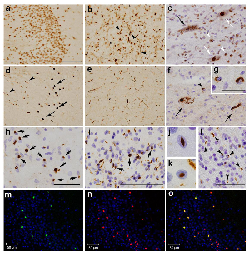
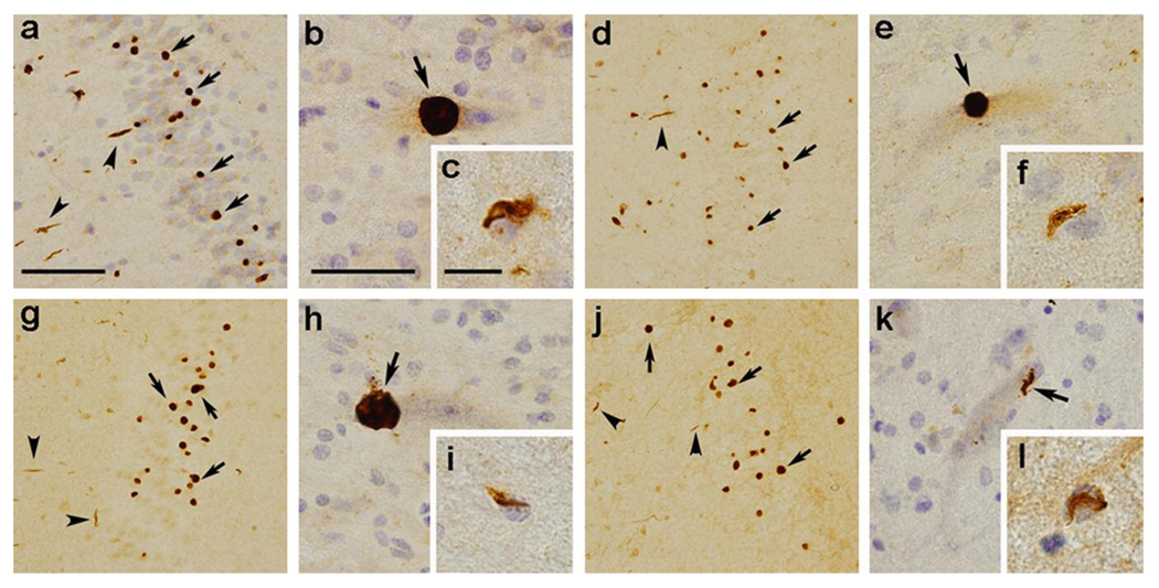
Objective: TAR DNA-binding protein of 43kDa (TDP-43) is deposited as cytoplasmic and intranuclear inclusions in brains of patients with frontotemporal lobar degeneration with ubiquitinated inclusions (FTLD-U) and amyotrophic lateral sclerosis (ALS). Previous studies reported that abnormal phosphorylation takes place in deposited TDP-43. The aim of this study was to identify the phosphorylation sites and responsible kinases, and to clarify the pathological significance of phosphorylation of TDP-43.

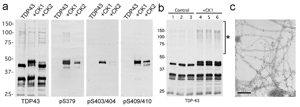
Methods: We generated multiple antibodies specific to phosphorylated TDP-43 by immunizing phosphopeptides of TDP-43, and analyzed FTLD-U and ALS brains by immunohistochemistry, immunoelectron microscopy, and immunoblots. In addition, we performed investigations aimed at identifying the responsible kinases, and we assessed the effects of phosphorylation on TDP-43 oligomerization and fibrillization.

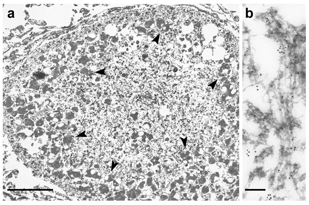
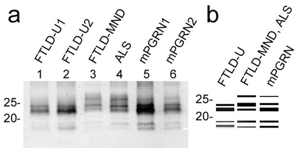
Results: We identified multiple phosphorylation sites in carboxyl-terminal regions of deposited TDP-43. Phosphorylation-specific antibodies stained more inclusions than antibodies to ubiquitin and, unlike existing commercially available anti-TDP-43 antibodies, did not stain normal nuclei. Ultrastructurally, these antibodies labeled abnormal fibers of 15nm diameter and on immunoblots recognized hyperphosphorylated TDP-43 at 45kDa, with additional 18 to 26kDa fragments in sarkosyl-insoluble fractions from FTLD-U and ALS brains. The phosphorylated epitopes were generated by casein kinase-1 and -2, and phosphorylation led to increased oligomerization and fibrillization of TDP-43.

Interpretation: These results suggest that phosphorylated TDP-43 is a major component of the inclusions, and that abnormal phosphorylation of TDP-43 is a critical step in the pathogenesis of FTLD-U and ALS. Phosphorylation-specific antibodies will be powerful tools for the investigation of these disorders.


Figures

Fig. 1
Fig. 1
Immunohistochemical comparison of FTLD-U and ALS brains using the phosphorylation independent anti-TDP-43 antibody (ProteinTech) (ac) and the phosphorylation dependent anti-TDP-43 antibody (pS409/410) (dl), in the dentate gyrus (a, d) and temporal cortex (b, e) of the sporadic FTLD-U cases, in the lumbar spinal cord (c, f, and g) and the frontal cortex (h) of the ALS cases and in the the frontal cortex (i, j, and k) and the frontal white matter (l) of the familial FTLD-U cases with PGRN mutations. a: Since most of the nuclei of dentate gyrus granular neurons are immunopositive with the phosphorylation-independent antibody, it is difficult to identify NCIs. b: TDP-43 positive DNs are recognizable (arrowheads) in addition to the nuclei. c: The black arrow indicates a cell with skein-like inclusions. White arrows and arrowheads indicate the normal nuclei of anterior horn cells and glial nuclei, respectively. Photomicrographs d ~ f illustrate the corresponding areas to a ~ c, respectively. Note the absence of nuclear staining in d ~ g with the phosphorylation-dependent antibody pS409/410. d: NCIs (arrows) and DNs (arrowheads) are clearly seen. e: More abundant DNs are seen than in b. f: Arrows indicate skein-like inclusions; the arrowheads indicate glial inclusions. The insert (g) shows glial inclusions at a higher magnification. h: NCIs in the frontal cortices of the ALS case are immunopositive. In the cases with PGRN mutations, pS409/410 clearly stains NCIs (arrows), DNs (i) and NIIs (j and k) in the superficial cortical layers, and abundant immunopositive structures in the white matter (arrowheads in l), with no nuclear staining. The sections are counterstained with hematoxylin to reveal nuclei in c, f, g, h, i, j, k, and l. Calibration bars a (also for b, d and e) and i indicate 100 µm; c (also for f), h and l indicate 50 µm; g denotes 25 µm; j (also for k) is 10 µm. mo: Anti-ubiquitin (DF2) and pS409/410 double-label immunofluorescence histochemistry of the dentate gyrus in the FTLD-U case. Only some of the pS409/410 positive NCIs are also ubiquitin positive. m DF2; n pS409/410; g merge. The cell nuclei are stained with TO-PRO-3 (Invitrogen, Tokyo, Japan), producing a blue color.
Fig. 2
Fig. 2
Immunohistochemistry of FTLD-U brains and ALS spinal cords using the phosphorylation-dependent anti-TDP-43 antibodies specific for pS379 (ac), pS403/404 (df), pS409 (gi), and pS410 (jl). These antibodies recognize NCIs (arrows in a, d, g, and j) and DNs (arrowheads in a, d, g, and j) in the dentate gyrus of the sporadic FTLD-U cases and motoneuronal round inclusions (arrow in b, e, and h), skein-like inclusion (arrow in k) and glial inclusions (c, f, i, and l) in the lumber spinal cord of the ALS cases. Note the absence of nuclear staining. The sections are counterstained with hematoxylin to reveal nuclei in a, b, c, e, f, h, i, k, and l. Bars a (also for d, g, and j) denote 100 µm; b (also for e, h, and k) is 25 µm; c (also for f, i, and l) is 12.5 µm.
Fig. 3
Fig. 3
a: A low power immunoelectron micrograph of a phosphorylated TDP-43-positive motoneuronal inclusion in the spinal cord of an ALS patient. The irregularly shaped structure surrounded by lipofuscins (arrowheads) is the inclusion. b: At higher magnification, abnormal filaments of 15 nm diameter are immunopositive. Immunoreaction with pS409/410, probed with immunogold particles (diameter 10 nm), appears as black dots. Bars indicate, for a 5 µm; b 500 nm.
Fig. 4
Fig. 4
a: Immunoblot analyses of sarkosyl-insoluble, urea-soluble fractions from control, AD, FTD (FTLD-U) and ALS brains with phosphorylation-independent anti-TDP-43 antibody (ProteinTech) (a) and phosphorylation-dependent anti-TDP-43 antibodies specific for pS409/410 (b), pS409 (c), pS410 (d), pS403/404 (e), and pS379 (f) before (−) and after (+) the treatment with lambda protein phosphatase (λPPase). a. With the phosphorylation-independent antibody, a positive band of 43 kDa is commonly seen in all cases (asterisk), while an additional band of 45 kDa is observed only in FTD and ALS (arrow), the labeling of which is abolished after dephosphorylation. bf. The phosphorylation-dependent antibodies specifically label the ~45 kDa band (arrow) and the ~25 kDa fragment (arrowhead) as well as a smear, only in FTD and ALS. These immunoreactivities are abolished after dephosphorylation. Normal 43 kDa TDP-43 in control and diseased brains is not stained by these phosphorylation-dependent antibodies. The two bands recognized by the antibody specific for pS403/404 in AD (double asterisk in e) disappear after dephosphorylation, suggesting a cross reaction of the antibody to other phosphorylated proteins.
Fig. 5
Fig. 5
A relationship between the clinicopathological subtypes of TDP-43 proteinopathies and the band pattern of the C-terminal fragments of phosphorylated TDP-43. a: Immunoblots of the sarkosyl-insoluble, urea-soluble fractions from sporadic FTLD-U, FTLD-MND, ALS and mPGRN cases with the pS409/410 antibody. The samples are loaded on 15% polyacrylamide gel. Sporadic FTLD-U cases (lanes 1, 2) show a band pattern with two major bands at 23 and 24 kDa and two minor bands at 18 and 19 kDa. A band of 24 kDa is weaker than that of 23 kDa, and a 19 kDa band is weaker than an 18 kDa band. FTLD-MND (lane 3) and ALS (lane 4) cases show a pattern with three major bands at 23, 24 and 26kDa and two minor bands at 18 and 19kDa. A 24 kDa band is the most intense, and an 18 kDa band is weaker than a 19 kDa band. mPGRN (lanes 5, 6) cases show three major bands at 23, 24 and 26kDa and two minor bands at 18 and 19kDa. A 23 kDa band is the most intense, and a band of 18 kDa and that of 19 kDa show similar intensity. The band pattern of mPGRN cases is therefore a composite of that seen in FTLD-U, FTLD-MND and ALS. b: Schematic diagram of the band pattern of the C-terminal fragments of phosphorylated TDP-43.
Fig. 6
Fig. 6
a: Immunoblot analyses of recombinant TDP-43 phosphorylated in vitro. The crude extract from E coli that expressed human TDP-43 is treated with CK1 and CK2 at 30°C for 14hrs, and probed with a phosphorylation-independent antibody against a C-terminal peptide of TDP-43 (405–414), and with phosphorylation-dependent antibodies pS379, pS403/404 and pS409/410. Phosphorylation by CK1 causes the mobility shift to ~45kDa and induction of intense immunoreactivity to the phosphorylation-dependent antibodies. b: Immunoblot analyses of recombinant TDP-43 phosphorylated by CK1. The recombinant TDP-43, which is partially purified by heparin-Toyopearl column chromatography, is incubated with (lanes 4~6) or without (lanes 1~3) CK1 in the presence of ATP at 37°C for 14 hrs, and probed with the phosphorylation-independent TDP-43 antibody (ProteinTech). Results in three independent, representative experiments are shown. Note the SDS-stable TDP-43 oligomers at ~100 ~ 200 kDa (asterisk) are detected after phosphorylation by CK1. c: Positive immunolabeling by pS409/410 of filaments assembled from recombinant TDP-43 phosphorylated by CK1 (10 nm colloidal gold). Bar denotes 200nm.

References

    1. Mackenzie IRA, Feldman HH. Ubiquitin immunohistochemistry suggests classic motor neuron disease, motor neuron disease with dementia, and frontotemporal dementia of the motor neuron disease type represent a clinicopathological spectrum. J Neuropathol Exp Neurol. 2005;64:730–739. - PubMed
    1. Baker M, Mackenzie IR, Pickering-Brown SM, et al. Mutations in progranulin cause tau-negative frontotemporal dementia linked to chromosome 17. Nature. 2006;442:916–919. - PubMed
    1. Cruts M, Gijselinck I, van der Zee J, et al. Null mutations in progranulin cause ubiquitin-positive frontotemporal dementia linked to chromosome 17q21. Nature. 2006;442:920–924. - PubMed
    1. Watts GDJ, Wymer J, Kovach MJ, et al. Inclusion body myopathy associated with Paget disease of bone and frontotemporal dementia is caused by mutant valosin-containing protein. Nat Genet. 2004;36:377–381. - PubMed
    1. Morita M, Al-Chalabi A, Anderson PM, et al. A locus on chromosome 9p confers susceptibility to ALS and frontotemporal dementia. Neurology. 2006;66:839–844. - PubMed

Publication types

MeSH terms