. 2008 Summer;36(2):305-14, 212.
doi: 10.1111/j.1748-720X.2008.00274.x.

The risks and benefits of searching for incidental findings in MRI research scans

Affiliations

The risks and benefits of searching for incidental findings in MRI research scans

Jason M Royal et al. J Law Med Ethics. 2008 Summer.

Abstract

We weigh the presumed benefits of routinely searching all research scans for incidental findings (IFs) against its substantial risks, including false-positive and false-negative findings, and the possibility of triggering unnecessary, costly evaluations and perhaps harmful treatments. We argue that routinely searching for IFs may not maximize benefits and minimize risks to participants.


Figures

Figure 1
Figure 1. Three Types of MRI Scans Commonly Used in Neuroimaging Research
(a) T1-weighted images, the most common in neuroimaging research, are used to investigate the anatomical structure of various regions of the brain. T1-weighted images can reveal only comparatively large, space-occupying tumors and hydrocephalus. The presence of a tumor appears in a T1-weighted image as a distortion of the anatomical structures that surround the space occupied by the tumor. Tumors themselves, however, cannot be seen. (b) T2-weighted images can reveal visually processes that are directly related to tumors, such as tissue edema and necrosis. However, this type of image is used far less frequently in neuroimaging research that is the T1-weighted image. (c) Echoplanar images, which are used in functional MRI, are of virtually no clinical value because of their extremely low resolution and poor signal-to-noise characteristics.
Figure 2
Figure 2. Incidental Findings (IFs) with Indeterminate Clinical Significance
(a) Unidentified bright objects (UBOs), which appear as bright spots on T2-weighted images (see yellow arrow in panel (a)), rarely can indicate the presence of vascular malformations, demyelinating plaques of multiple sclerosis, infarctions, or other entities. They also can appear as nonspecific, clinically indeterminate IFs, however, primarily in older populations, as well as in younger populations with various neurological or psychiatric illnesses. (b) Ventriculomegaly, or enlargement of the ventricles, is a nonspecific finding that has numerous possible causes, including destruction of tissue around the ventricles or obstruction of the flow of cerebrospinal fluid. (c) Chiari I malformation is a condition in which a portion of the cerebellum (the cerebellar tonsils in particular) has dropped below the protective encasing of the skull (through the foramen magnum), a process known as “ectopia” (see yellow arrow in panel (c)). Symptoms usually appear in midlife, but Chiari I malformations and ectopia often are identified as IFs in adolescent populations in neuroimaging studies. Severe ectopia can put an individual at risk for potentially life-threatening injury to the cervical spinal cord, which regulates a number of vital bodily functions. The clinical significance of mild or borderline ectopia, however, is unclear, and individuals with mild ectopia usually remain entirely asymptomatic. The identification of such IFs with indeterminate clinical significance can expose participants to the burden and risks of treatment or ongoing clinical monitoring, which may be unnecessary and of little clinical benefit.
Figure 3
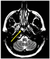
Figure 3. Limitations of Research-Quality Scans Can Increase Risk and Burden to Participants
A radiologist identified in the T2-weighted brain scan of this 49-year-old woman an abnormality that appears as a bright spot (a “hyperintensity”) located inferior to the sphenoid sinus and superior to the nasopharynx (indicated by an arrow). The abnormality initially was thought most likely to be a benign and relatively common type of cyst (termed a “Thornwaldt” cyst). This diagnosis could not be confirmed, however, based on a reading of our research-quality scans. Because of the outside possibility that the anomaly could have been a tumor or some other worrisome lesion, the participant was referred for subsequent clinical examination and MRI. Further consultation revealed no evidence of a dangerous tumor or lesion. The participant nevertheless was advised to undergo a course of periodic examination and additional MRIs — a “wait and watch” approach. In this instance, the limited information in our research-quality scan, compared with the more conclusive information that a detailed and definitive clinical scan provided, drew the participant into an open-ended program of clinical monitoring that may be neither necessary nor beneficial.

References

    1. Davis FG, et al. Prevalence Estimates for Primary Brain Tumors in the United States by Behavior and Major Histology Groups. Neuro-Oncology. 2001;3(3):152–158. - PMC - PubMed
    1. Rinkel GJ, et al. Prevalence and Risk of Rupture of Intracranial Aneurysms: A Systematic Review. Stroke. 1998;29(1):251–256. - PubMed
    2. Wardlaw JM, White PM. The Detection and Management of Unruptured Intracranial Aneurysms. Brain. 2000;123(Pt. 2):205–221. - PubMed
    1. Lee T, et al. Aggregate Analysis of the Literature for Unruptured Intracranial Aneurysm Treatment. American Journal of Neuroradiology. 2005;26(8):1902–1908. - PMC - PubMed
    1. Meadows J, et al. Asymptomatic Chiari Type I Malformations Identified on Magnetic Resonance Imaging. Journal of Neurosurgery. 2000;92(6):920–926. - PubMed
    1. Illes J, et al. Ethical Consideration of Incidental Findings on Adult Brain MRI in Research. Neurology. 2004;62(6):888–890. - PMC - PubMed
    2. Katzman GL, et al. Incidental Findings on Brain Magnetic Resonance Imaging from 1000 Asymptomatic Volunteers. JAMA. 1999;282(1):36–39. - PubMed
    3. Kim BS, et al. Incidental Findings on Pediatric MR Images of the Brain. American Journal of Neuroradiology. 2002;23(10):1674–1677. - PMC - PubMed
    4. Kumra S, et al. Ethical and Practical Considerations in the Management of Incidental Findings in Pediatric MRI Studies. Journal of the American Academy of Child and Adolescent Psychiatry. 2006;45(8):1000–1006. - PubMed
    5. Weber F, Knopf H. Incidental Findings in Magnetic Resonance Imaging of the Brains of Healthy Young Men. Journal of the Neurological Sciences. 2006;240(1-2):81–84. - PubMed

Publication types