Comparative Study
. 2008 Aug;146(2):298-309.
doi: 10.1016/j.ajo.2008.04.012. Epub 2008 Jun 10.

Retinopathy of prematurity management using single-image vs multiple-image telemedicine examinations

Affiliations
Comparative Study

Retinopathy of prematurity management using single-image vs multiple-image telemedicine examinations

Alexandra Lajoie et al. Am J Ophthalmol. 2008 Aug.

Abstract

Purpose: To compare performance of single-image vs multiple-image telemedicine examinations for retinopathy of prematurity (ROP) diagnosis.

Design: Prospective comparative study.

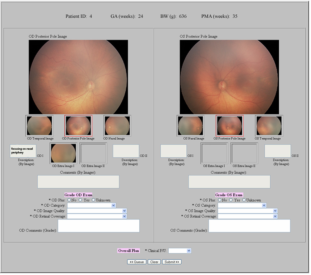
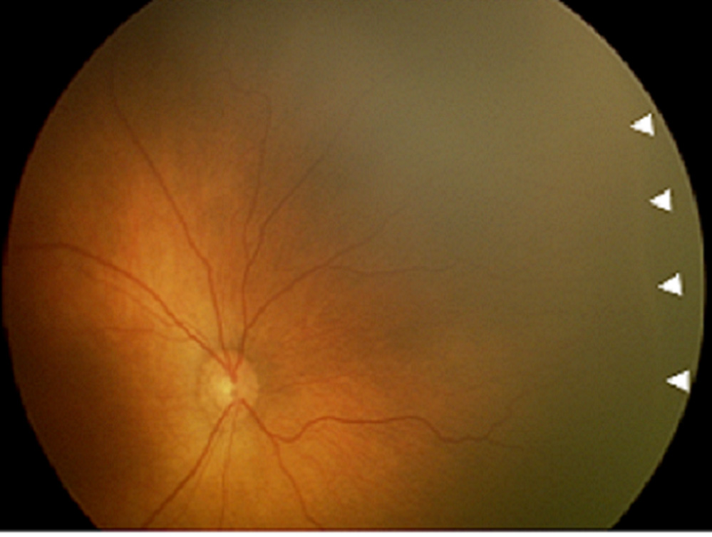
Methods: A total of 248 eyes from 67 consecutive infants underwent wide-angle retinal imaging by a trained neonatal nurse at 31 to 33 weeks and/or 35 to 37 weeks postmenstrual age (PMA) at a single academic institution. Data were uploaded to a web-based telemedicine system and interpreted by three masked retinal specialists. Diagnoses were provided based on single images, and subsequently on multiple images, from both eyes of each infant. Findings were compared to a reference standard of indirect ophthalmoscopy by a pediatric ophthalmologist. Primary outcome measures were recommended follow-up interval, presence of plus disease, presence of type-2 or worse ROP, and presence of visible peripheral ROP.

Results: Among the three graders, mean sensitivity/specificity for detection of infants requiring follow-up in less than one week were 0.85/0.93 by single-image examination and 0.91/0.88 by multiple-image examination at 35 to 37 weeks PMA. Mean sensitivity/specificity for detection of infants with type-2 or worse ROP were 0.82/0.95 by single-image examination and 1.00/0.91 by multiple-image examination at 35 to 37 weeks PMA. Mean sensitivity/specificity for detection of plus disease were 1.00/0.86 by single-image examination and 1.00/0.87 by multiple-image examination at 35 to 37 weeks PMA. There were no statistically-significant intragrader differences between accuracy of single-image and multiple-image telemedicine examinations for detection of plus disease.

Conclusions: Single-image and multiple-image telemedicine examinations perform comparably for determination of recommended follow-up interval and detection of plus disease. This may have implications for development of screening protocols, particularly in areas with limited access to ophthalmic care.


Figures

Figure 1
Figure 1. Web-based telemedicine system for single-image examinations for retinopathy of prematurity (ROP)
A wide-angle posterior image of each retina is displayed for graders.
Figure 2
Figure 2. Web-based telemedicine system for multiple-image examinations for retinopathy of prematurity (ROP)
Wide-angle posterior, nasal, and temporal images of each retina are displayed for graders, along with up to 2 additional images per eye if felt by nurse photographer to be of diagnostic value.
Figure 3
Figure 3. Graphical comparison of recommended follow-up intervals by single-image, multiple-image, and ophthalmoscopic examinations for retinopathy of prematurity
Exams were performed at 31–33 weeks and 35–37 weeks post-menstrual age (PMA). Results are displayed as percentage of infants, for single-image and multiple-image examinations vs. ophthalmoscopic examinations. *At both 31–33 weeks and 35–37 weeks PMA, there were no significant differences between the “single-image exam vs. ophthalmoscopy” follow-up interval distribution and the “multiple-image exam vs. ophthalmoscopy” follow-up distribution.
Figure 4
Figure 4. Graphical comparison of recommended follow-up intervals by single-image vs. multiple-image telemedicine examinations for retinopathy of prematurity
Exams were performed at 31–33 weeks and 35–37 weeks post-menstrual age (PMA). Results are displayed as percentage of infants. *At both 31–33 weeks and 35–37 weeks PMA, there was a significant difference (p<0.001) between the “single-image exam vs. multiple-image exam” follow-up distribution and those shown in Figure 3 (“single-image exam vs. ophthalmoscopy” and “multiple-image exam vs. ophthalmoscopy”).
Figure 5
Figure 5. Example of diagnostic disagreement by single-image telemedicine examination for retinopathy of prematurity (ROP), compared to multiple-image and ophthalmoscopic examinations
This was diagnosed as “no visible peripheral ROP” from single-image exam ([left] image) by all graders, as “mild ROP” from multiple-image exam ([left] and [right] images, plus temporal view) by all graders, and as “mild ROP” from ophthalmoscopic exam. Arrowheads show location of disease identified by graders.
Figure 6
Figure 6. Example of diagnostic agreement by single-image telemedicine examination for retinopathy of prematurity (ROP), compared to multiple-image and ophthalmoscopic examinations
This was diagnosed as “type-2 or worse ROP” from single-image exam by all graders, as “treatment-requiring ROP” from multiple-image exam (this image, plus temporal and nasal views) by all graders, and as “type-2 prethreshold ROP” from ophthalmoscopic exam. Arrowheads show location of disease identified by graders.

Similar articles

Cited by

References

    1. Cryotherapy for Retinopathy of Prematurity Cooperative Group. Multicenter trial of cryotherapy for retinopathy of prematurity: preliminary results. Arch Ophthalmol. 1988;106:471–479. - PubMed
    1. Early Treatment for Retinopathy of Prematurity Cooperative Group. Revised indications for the treatment of retinopathy of prematurity: results of the early treatment for retinopathy of prematurity randomized trial. Arch Ophthalmol. 2003;121:1684–1694. - PubMed
    1. Gilbert C, Foster A. Childhood blindness in the context of VISION 2020: the right to sight. Bull World Health Organ. 2001;79:227–232. - PMC - PubMed
    1. Gilbert C, Fielder A, Gordillo L, et al. Characteristics of infants with severe retinopathy of prematurity in countries with low, moderate, and high levels of development: Implications for screening programs. Pediatrics. 2005;115:e518–e525. - PubMed
    1. American Academy of Ophthalmology. Ophthalmologists warn of shortage in specialists who treat premature babies with blinding eye condition. [Accessed February 29, 2008]. Available at http://www.aao.org/newsroom/release/20060713.cfm.

Publication types

MeSH terms