. 2010 Mar;31(3):398-408.
doi: 10.1016/j.neurobiolaging.2008.05.010. Epub 2008 Jun 10.

Deregulation of sphingolipid metabolism in Alzheimer's disease

Affiliations

Deregulation of sphingolipid metabolism in Alzheimer's disease

Xingxuan He et al. Neurobiol Aging. 2010 Mar.

Abstract

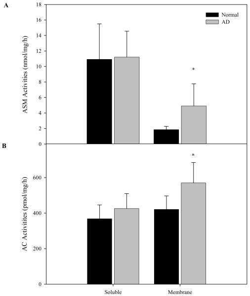
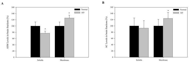
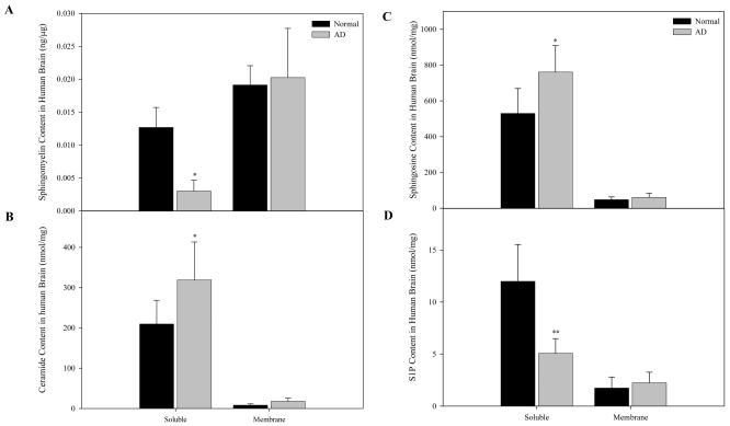
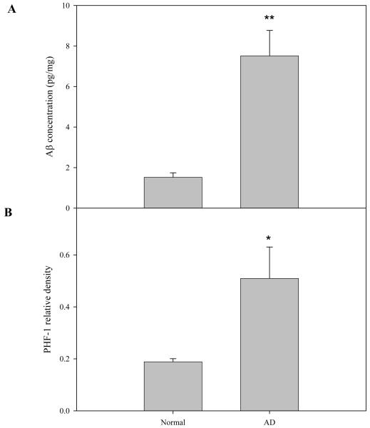
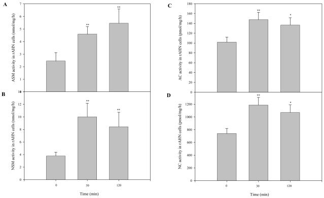
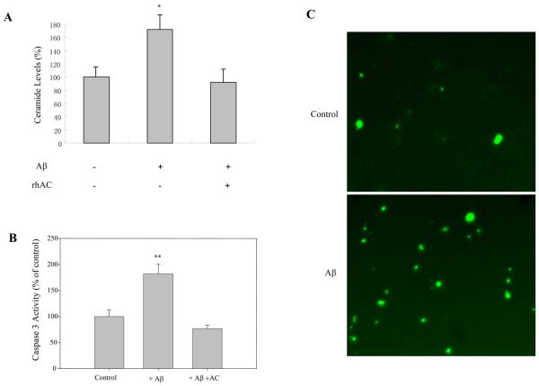
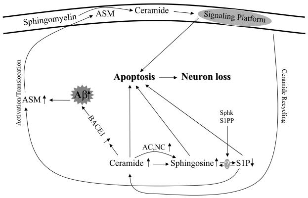
Abnormal sphingolipid metabolism has been previously reported in Alzheimer's disease (AD). To extend these findings, several sphingolipids and sphingolipid hydrolases were analyzed in brain samples from AD patients and age-matched normal individuals. We found a pattern of elevated acid sphingomyelinase (ASM) and acid ceramidase (AC) expression in AD, leading to a reduction in sphingomyelin and elevation of ceramide. More sphingosine also was found in the AD brains, although sphingosine-1-phosphate (S1P) levels were reduced. Notably, significant correlations were observed between the brain ASM and S1P levels and the levels of amyloid beta (Abeta) peptide and hyperphosphorylated tau protein. Based on these findings, neuronal cell cultures were treated with Abeta oligomers, which were found to activate ASM, increase ceramide, and induce apoptosis. Pre-treatment of the neurons with purified, recombinant AC prevented the cells from undergoing Abeta-induced apoptosis. We propose that ASM activation is an important pathological event leading to AD, perhaps due to Abeta deposition. The downstream consequences of ASM activation are elevated ceramide, activation of ceramidases, and production of sphingosine. The reduced levels of S1P in the AD brain, together with elevated ceramide, likely contribute to the disease pathogenesis.


Figures

Fig. 1
Fig. 1. ASM and AC activities in normal and AD brain
Soluble (cytosolic) and membrane fractions from normal (N=6) and AD (N=9) brains were prepared as described in the “Materials and Methods”. ASM and AC activities were measured after 1 hr and overnight incubations at 37°C, respectively. *p<0.05, compared to normal brains. Values are expressed as the mean ± S.D.
Fig. 2
Fig. 2. ASM and AC protein levels in normal and AD brain
Soluble (cytosolic) and membrane fractions from normal (N=6) and AD (N=9) brains were prepared as described in the “Materials and Methods”. Rabbit polyclonal antibodies against human ASM and AC were used for western blotting as described. The ASM and AC-specific protein bands were scanned and the signals were normalized to β-actin signals from the same blot (see “Materials and Methods”). *p<0.05, compared to normal brains. Values are expressed as the percentage of control.
Fig. 3
Fig. 3. Sphingomyelin, ceramide, sphingosine, and S1P contents of normal and AD brain
Soluble (cytosolic) and membrane fractions from normal (N=6) and AD (N=9) brains were prepared as described in the “Materials and Methods”. For each fraction two lipid extracts were prepared, one containing S1P and the other containing sphingomyelin, ceramide and sphingosine. Sphingomyelin, ceramide, sphingosine, and S1P were determined using HPLC based methods as described in the “Materials and Methods”. *p<0.05, **p<0.01, compared to normal brains. Values are expressed as the mean ± S.D.
Fig. 4
Fig. 4. Aβ and PHF-1 levels in normal and AD brain
Soluble (cytosolic) fractions from normal (N=6) and AD (N=9) brains were prepared as described in the “Materials and Methods”. Aβ levels in these fractions were determined using a human Aβ42 ELISA kit (see “Materials and Methods”). Mouse monoclonal antibody against human hyperphosphorylated tan protein, PHF-1, was used for western blotting as described. The PHF-1 protein bands were scanned and the signals were normalized to β-actin signals from the same blot. *p<0.05, **p<0.01, compared to normal brains. Values are expressed as the mean ± S.D.
Fig. 5
Fig. 5. Sphingomyelinase and ceramidase activities in neuronal cultures after Aβ treatment
After 5-7 days of growth in differentiation media, neuronal cultures were treated with 1 μM of Aβ o1□□□□ers for the indicated times. Cell lysates were then prepared and sphingomyelinase and ceramidase activities were measured after incubation at 37°C for 1 hr and overnight, respectively. *p<0.05, **p<0.01, compared to normal brains. Values are expressed as the mean ± S.D (N=3). A, ASM and NSM activities. B, AC and NC activities.
Fig. 6
Fig. 6. The effect of Aβ on ceramide levels and apoptosis in neuronal cultures
After 3-5 days of growth in differentiation media, neuronal cultures were treated with 1 μM of Aβ for 30 min with or without 1 hr of rhAC pre-treatment (1 μg/ml). Ceramide levels (A) were verified using the DAG kinase method (He et al., 2001). Caspase 3 activity (B) was measured using EnzCheck Caspase-3 assay kit. *p<0.05, **p<0.01, compared to normal brains. Values are expressed as the mean ± S.D (N=3). C, Neuronal cultures were treated with 1 μM of Aβ for 17 hr, and then processed with the DeadEnd Fluorometric TUNEL system and analyzed by microscopy. Pictures were captured in the green channel (20x magnification).
Fig. 7
Fig. 7. Schematic pathway proposed for the Aβ-induced sphingolipid changes in the AD brain
Sphk, sphingosine kinase, S1PP, sphingosine-1-phosphate phosphatase, Lyase, sphingosine-1-phosphate lyase.

References

    1. Alessenko AV, Bugrova AE, Dudnik LB. Connection of lipid peroxide oxidation with the sphingomyelin pathway in the development of Alzheimer's disease. Biochem. Soc. Trans. 2004;32:144–146. - PubMed
    1. Ayasolla K, Khan M, Singh AK, Singh I. Inflammatory mediator and beta-amyloid (25-35)-induced ceramide generation and iNOS expression are inhibited by vitamin E. Free Radic. Biol. Med. 2004;37:325–338. - PubMed
    1. Bollinger CR, Teichgraber V, Gulbins E. Ceramide-enriched membrane domains. Biochim. Biophys. Acta. 2005;1746:284–294. - PubMed
    1. Buchet R, Pikula S. Alzheimer's disease: its origin at the membrane, evidence and questions. Acta Biochim. Pol. 2000;47:725–733. - PubMed
    1. Chen H, Tung YC, Li B, Iqbal K, Grundke-Iqbal I. Trophic factors counteract elevated FGF-2-induced inhibition of adult neurogenesis. Neurobiol. Aging. 2007;28:1148–1162. - PubMed

Publication types

MeSH terms