. 2008 Jun 12;453(7197):925-9.
doi: 10.1038/nature07055.

Substrate-targeting gamma-secretase modulators

Affiliations

Substrate-targeting gamma-secretase modulators

Thomas L Kukar et al. Nature. .

Abstract

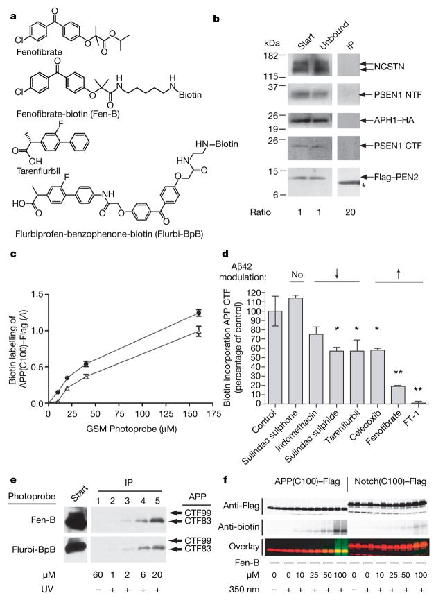
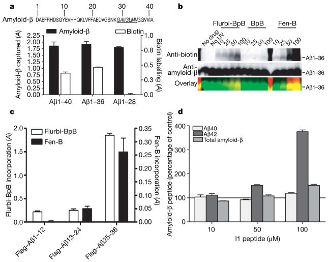
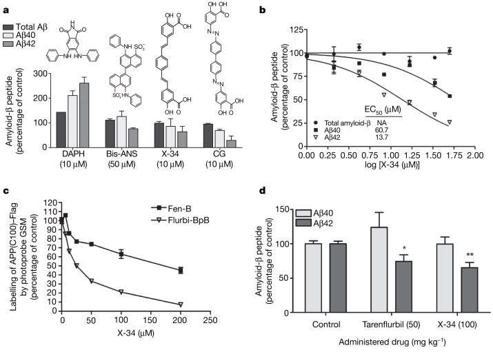
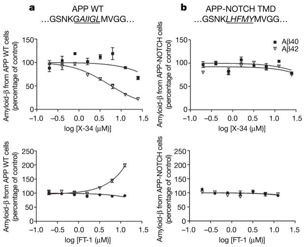
Selective lowering of Abeta42 levels (the 42-residue isoform of the amyloid-beta peptide) with small-molecule gamma-secretase modulators (GSMs), such as some non-steroidal anti-inflammatory drugs, is a promising therapeutic approach for Alzheimer's disease. To identify the target of these agents we developed biotinylated photoactivatable GSMs. GSM photoprobes did not label the core proteins of the gamma-secretase complex, but instead labelled the beta-amyloid precursor protein (APP), APP carboxy-terminal fragments and amyloid-beta peptide in human neuroglioma H4 cells. Substrate labelling was competed by other GSMs, and labelling of an APP gamma-secretase substrate was more efficient than a Notch substrate. GSM interaction was localized to residues 28-36 of amyloid-beta, a region critical for aggregation. We also demonstrate that compounds known to interact with this region of amyloid-beta act as GSMs, and some GSMs alter the production of cell-derived amyloid-beta oligomers. Furthermore, mutation of the GSM binding site in the APP alters the sensitivity of the substrate to GSMs. These findings indicate that substrate targeting by GSMs mechanistically links two therapeutic actions: alteration in Abeta42 production and inhibition of amyloid-beta aggregation, which may synergistically reduce amyloid-beta deposition in Alzheimer's disease. These data also demonstrate the existence and feasibility of 'substrate targeting' by small-molecule effectors of proteolytic enzymes, which if generally applicable may significantly broaden the current notion of 'druggable' targets.


Figures

Figure 1
Figure 1. GSM photoprobes label APP CTF
a, Structures of the parent GSMs (fenofibrate and tarenflurbil) and photoprobe derivates (Fen-B and Flurbi-BpB) are shown. b, The absence of PSEN1, NCSTN, APH1 and PEN2 labelling by the GSM Fen-B in a purified γ-secretase preparation (from CHO γ-30 cells12) and immunoprecipitation with streptavidin. The ratios of sample relative to the starting material are shown. Start and unbound lanes contain 5% of the immunoprecipitated material (lane 3), therefore the ratios are 1, 1 and 20. Asterisk denotes nonspecific reactivity with streptavidin. c, GSM photoprobes (Flurbi-BpB, closed circles, and Fen-B, open triangles) label a recombinant APP γ-secretase substrate (APP(C100)–Flag) with similar potency. A, absorbance; data are mean ± s.e.m., n = 2. d, Labelling of APP(C100)–Flag by Fen-B (10 μM) is competed by Aβ42-lowering and -raising GSMs (100 μM) but not by sulindac sulphone, a non-GSM NSAID. Data are presented as percentage control ±s.e.m., n = 2. Asterisk, P <0.05; two asterisks, P <0.01; ANOVA with Dunnett’s post-hoc analysis. e, GSM photoprobes label APP CTF from cells. CHAPSO solubilized membrane fractions from H4 APP-CTF–alkaline phosphatase cells were crosslinked with Fen-B and Flurbi-BpB (50 μM) and analysed by immunoprecipitation with streptavidin and immunblotting for APP (antibody CT20). Both GSMs label a fragment of APP that co-migrates with APP(C83). UV, ultraviolet. f, A GSM photoprobe preferentially labels a recombinant APP substrate (APP(C100)–Flag; left panel) relative to Notch (Notch(C100)–Flag; right panel). Samples were analysed by western blotting for incorporation of Fen-B. Green, biotin; red, Flag; yellow, dual reactivity; LiCor Odyssey.
Figure 2
Figure 2. GSM photoprobes bind to the amyloid-β region of APP
a, Fen-B labels Aβ1–40 and Aβ1–36 but not Aβ1–28, suggesting that the binding site for Fen-B is located between residues 28 to 36 of amyloid-β, which are highlighted in italics. b, Flurbi-BpB and Fen-B label Aβ1–36 (biotin incorporation), whereas the photoaffinity tag alone (BpB) shows minor labelling. c, Flurbi-BpB and Fen-B preferentially label Flag-tagged Aβ25–36. Data are presented as biotin incorporation (absorbance, A) ±s.e.m., n = 3. d, The peptide fragment I1 (NH2-FEGKF-CONH2) increases Aβ42 in H4 cells expressing APP similar to the GSM fenofibrate.
Figure 3
Figure 3. Compounds that bind Aβ are GSMs in vitro and in vivo
a, A cell-based screen of Aβ-binders identified molecules that increase Aβ42 (DAPH) or decrease Aβ42 (Bis-ANS, X-34 or chrysamine G (CG)). Data are mean ± s.e.m., n = 3. b, X-34 is an Aβ42-lowering GSM. Changes in amyloid-β peptide amounts after X-34 treatment are shown. Data are presented as percentage control ±s.e.m., n = 3. EC50 values were calculated as described in Methods. Total amyloid-β did not decrease. NA, not applicable. c, X-34 binds to APP(C100)–Flag and decreases labelling by GSM photoprobes. Biotin incorporation into APP by Fen-B and Flurbi-BpB is presented as percentage of control (peptide without X-34) ±s.e.m., n = 2. d, X-34 lowers Aβ42 in Tg2576 mice after 4 h. X-34 (n = 7) and tarenflurbil (n = 5) reduce Aβ42 selectively; control (n = 7). Data are presented as amyloid-β percentage of control relative to vehicle ±s.e.m. Animals per group (n). Asterisk, P <0.05; two asterisks, P <0.01; ANOVA with Dunnett’s post-hoc analysis.
Figure 4
Figure 4. The ability of GSMs to shift Aβ42 amounts is sensitive to the amino acid sequence of the binding site on APP
a, b, The sequences of wild-type (WT; a) APP and the mutated substrate (b) containing the homologous region (italic, underlined) of the NOTCH transmembrane domain (TMD) in APP where GSMs are hypothesized to bind. Top row, X-34 lowers Aβ42 (EC50 5.9 μM) from APP wild-type cells (a) but did not change either Aβ40 or Aβ42 concentrations in the APP-NOTCH TMD line (b). Bottom row, FT-1 (12.5 μM) raised Aβ42 200% in APP wild-type cells (a); however, in APP-NOTCH TMD cells (b), FT-1 caused minimal changes in the Aβ42 (95%) or Aβ40 (90%) signal. Data are presented as amyloid-β percentage of control ±s.e.m., n= 3.

Comment in

References

    1. Kukar T, Golde TE. Possible mechanisms of action of NSAIDs and related compounds that modulate γ-secretase cleavage. Curr Top Med Chem. 2008;8:47–53. - PMC - PubMed
    1. Kodadek T. Inhibition of proteolysis and other posttranslational modifications with substrate-targeted inhibitors. Biopolymers. 2002;66:134–140. - PubMed
    1. Eriksen JL, et al. NSAIDs and enantiomers of flurbiprofen target γ-secretase and lower Aβ 42 in vivo. J Clin Invest. 2003;112:440–449. - PMC - PubMed
    1. Weggen S, et al. A subset of NSAIDs lower amyloidogenic Aβ42 independently of cyclooxygenase activity. Nature. 2001;414:212–216. - PubMed
    1. Christensen DD. Changing the course of Alzheimer’s disease: anti-amyloid disease-modifying treatments on the horizon. Prim Care Companion J Clin Psychiatry. 2007;9:32–41. - PMC - PubMed

Publication types

MeSH terms

Substances