

Pontin is localized in nucleolar fibrillar centers
- PMID: 18548265
- PMCID: PMC2564108
- DOI: 10.1007/s00412-008-0170-8
Pontin is localized in nucleolar fibrillar centers
Abstract
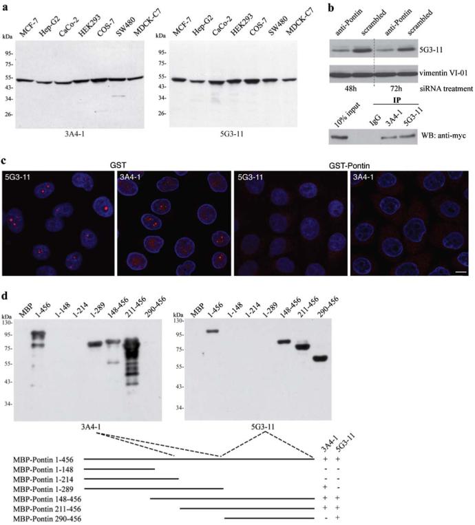
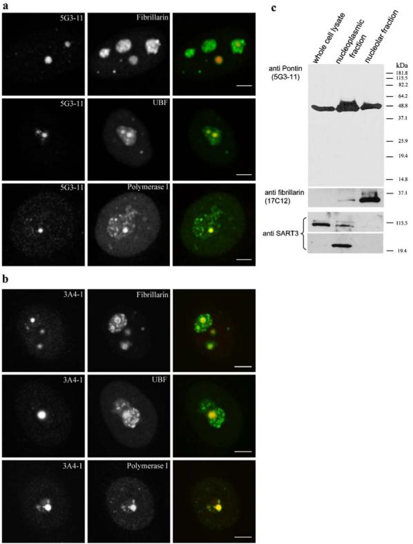
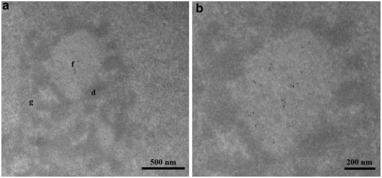
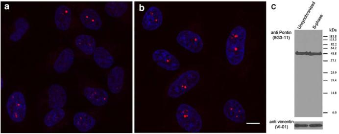
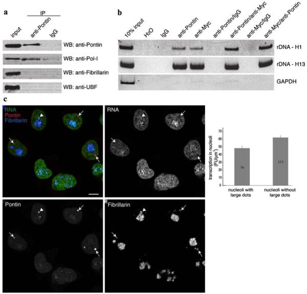
Pontin is a multifunctional protein having roles in various cellular processes including regulation of gene expression. Here, we addressed Pontin intracellular localization using two different monoclonal antibodies directed against different Pontin epitopes. For the first time, Pontin was directly visualized in nucleoli where it co-localizes with Upstream Binding Factor and RNA polymerase I. Nucleolar localization of Pontin was confirmed by its detection in nucleolar extracts and by electron microscopy, which revealed Pontin accumulation specifically in the nucleolar fibrillar centers. Pontin localization in the nucleolus was dynamic and Pontin accumulated in large nucleolar dots mainly during S-phase. Pontin concentration in the large nucleolar dots correlated with reduced transcriptional activity of nucleoli. In addition, Pontin was found to associate with RNA polymerase I and to interact in a complex with c-Myc with rDNA sequences indicating that Pontin is involved in the c-Myc-dependent regulation of rRNA synthesis.
Figures
Similar articles
-
Nucleolar localization of upstream binding factor in HeLa cells depends on rRNA synthesis activities.Folia Biol (Praha). 2008;54(6):202-6. Folia Biol (Praha). 2008. PMID: 19393134
-
Does the synthesis of ribosomal RNA take place within nucleolar fibrillar centers or dense fibrillar components? A critical appraisal.J Struct Biol. 1995 Jan-Feb;114(1):1-22. doi: 10.1006/jsbi.1995.1001. J Struct Biol. 1995. PMID: 7772414
-
Nucleolar ultrastructure and protein allocation in in vitro produced porcine embryos.Mol Reprod Dev. 2004 Jul;68(3):327-34. doi: 10.1002/mrd.20088. Mol Reprod Dev. 2004. PMID: 15112326
-
Nucleolar localization of murine nuclear DNA helicase II (RNA helicase A).J Cell Sci. 1999 Aug;112 ( Pt 16):2693-703. doi: 10.1242/jcs.112.16.2693. J Cell Sci. 1999. PMID: 10413677
-
The nucleolus.Anat Embryol (Berl). 1993 Dec;188(6):515-36. doi: 10.1007/BF00187008. Anat Embryol (Berl). 1993. PMID: 8129175 Review.
Cited by
-
Functional ultrastructure of the plant nucleolus.Protoplasma. 2014 Nov;251(6):1285-306. doi: 10.1007/s00709-014-0648-6. Epub 2014 Apr 23. Protoplasma. 2014. PMID: 24756369 Free PMC article. Review.
-
A Rapid Approach for Identifying Cell Lines Lacking Functional Cytidine Deaminase.Int J Mol Sci. 2025 Apr 3;26(7):3344. doi: 10.3390/ijms26073344. Int J Mol Sci. 2025. PMID: 40244204 Free PMC article.
-
The emergence of the conserved AAA+ ATPases Pontin and Reptin on the signaling landscape.Sci Signal. 2013 Mar 12;6(266):mr1. doi: 10.1126/scisignal.2003906. Sci Signal. 2013. PMID: 23482663 Free PMC article.
-
Structure and epigenetics of nucleoli in comparison with non-nucleolar compartments.J Histochem Cytochem. 2010 May;58(5):391-403. doi: 10.1369/jhc.2009.955435. Epub 2009 Dec 21. J Histochem Cytochem. 2010. PMID: 20026667 Free PMC article. Review.
-
The yeast prefoldin-like URI-orthologue Bud27 associates with the RSC nucleosome remodeler and modulates transcription.Nucleic Acids Res. 2014 Sep;42(15):9666-76. doi: 10.1093/nar/gku685. Epub 2014 Jul 31. Nucleic Acids Res. 2014. PMID: 25081216 Free PMC article.
References
-
- Andersen JS, Lyon CE, Fox AH, Leung AK, Lam YW, Steen H, Mann M, Lamond AI. Directed proteomic analysis of the human nucleolus. Curr Biol. 2002;12:1–11. - PubMed
-
- Arabi A, Wu S, Ridderstrale K, Bierhoff H, Shiue C, Fatyol K, Fahlen S, Hydbring P, Soderberg O, Grummt I, Larsson LG, Wright AP. c-Myc associates with ribosomal DNA and activates RNA polymerase I transcription. Nat Cell Biol. 2005;7:303–310. - PubMed
Publication types
MeSH terms
Substances
Grants and funding
LinkOut - more resources
Full Text Sources
Miscellaneous
