. 2008 Jul 8;47(27):7304-16.
doi: 10.1021/bi800088t. Epub 2008 Jun 13.

A second conserved GAF domain cysteine is required for the blue/green photoreversibility of cyanobacteriochrome Tlr0924 from Thermosynechococcus elongatus

Affiliations

A second conserved GAF domain cysteine is required for the blue/green photoreversibility of cyanobacteriochrome Tlr0924 from Thermosynechococcus elongatus

Nathan C Rockwell et al. Biochemistry. .

Abstract

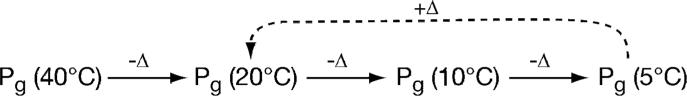
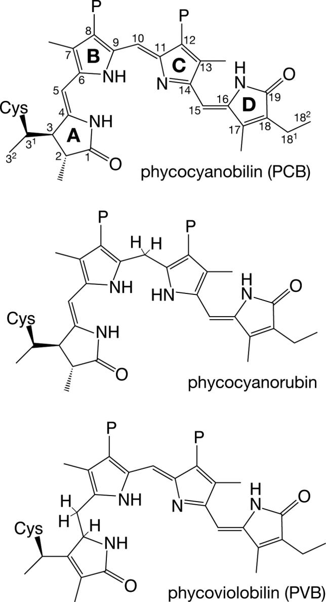
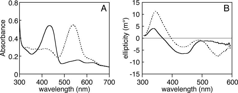
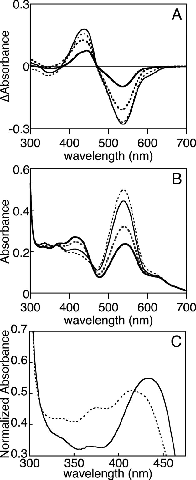
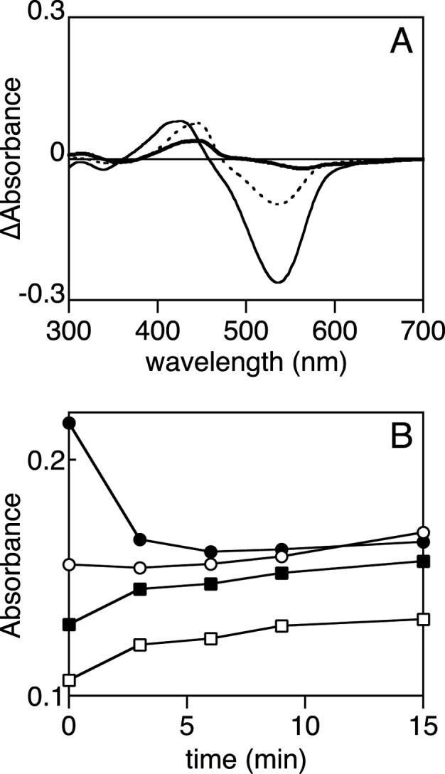
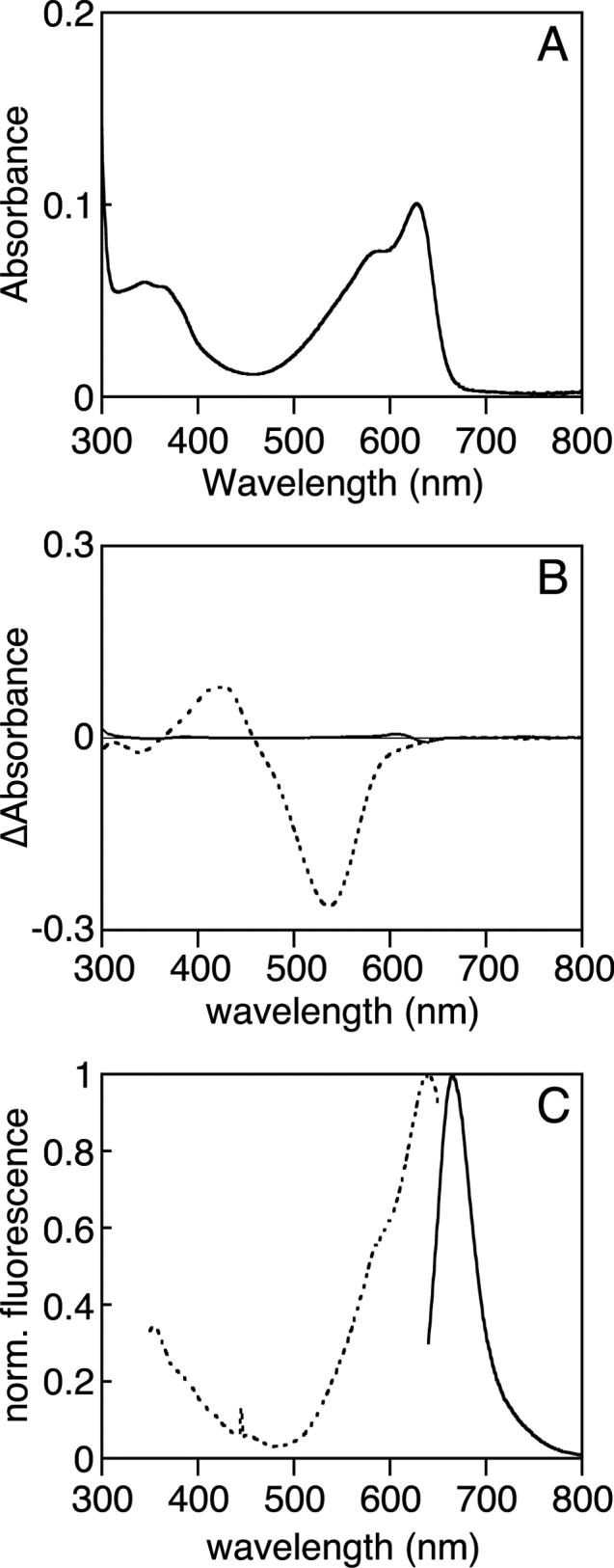
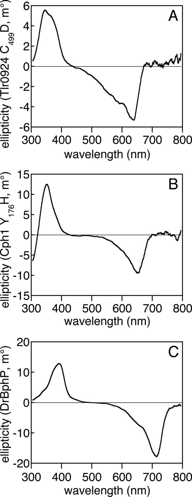
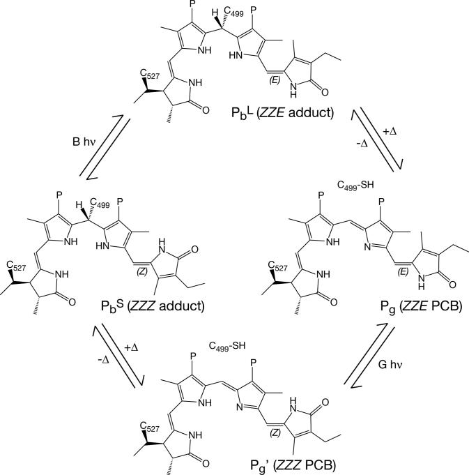
Phytochromes are widely occurring red/far-red photoreceptors that utilize a linear tetrapyrrole (bilin) chromophore covalently bound within a knotted PAS-GAF domain pair. Cyanobacteria also contain more distant relatives of phytochromes that lack this knot, such as the phytochrome-related cyanobacteriochromes implicated to function as blue/green switchable photoreceptors. In this study, we characterize the cyanobacteriochrome Tlr0924 from the thermophilic cyanobacterium Thermosynechococcus elongatus. Full-length Tlr0924 exhibits blue/green photoconversion across a broad range of temperatures, including physiologically relevant temperatures for this organism. Spectroscopic characterization of Tlr0924 demonstrates that its green-absorbing state is in equilibrium with a labile, spectrally distinct blue-absorbing species. The photochemically generated blue-absorbing state is in equilibrium with another species absorbing at longer wavelengths, giving a total of 4 states. Cys499 is essential for this behavior, because mutagenesis of this residue results in red-absorbing mutant biliproteins. Characterization of the C 499D mutant protein by absorbance and CD spectroscopy supports the conclusion that its bilin chromophore adopts a similar conformation to the red-light-absorbing P r form of phytochrome. We propose a model photocycle in which Z/ E photoisomerization of the 15/16 bond modulates formation of a reversible thioether linkage between Cys499 and C10 of the chromophore, providing the basis for the blue/green switching of cyanobacteriochromes.


Figures

Scheme 1
Scheme 1
Figure 1
Figure 1
Thioether adducts of linear tetrapyrroles. Top, the cyanobacterial phytochrome Cph1 utilizes phycocyanobilin (PCB) as chromophore. Cys259 forms a covalent linkage with the C31 atom of PCB (8, 18). PixJ is known to form a similar covalent linkage to PCB (24). Ring designations are indicated in bold, and the numbering for the carbon atoms is indicated. Middle, phycocyanorubin can be produced from PCB by reduction of the C10 position. This can be accomplished enzymatically with isolated PCB (66) or can be performed with chemical reductants for denatured phycobiliproteins (65). Bottom, α-phycoerythrocyanin (α-PEC) utilizes PCB as chromophore precursor, but PCB is tautomerized to phycoviolobilin (PVB) during the assembly reaction by accessory factors (79, 80). The tautomerization of PCB to PVB results in a shorter conjugated system with a blue-shifted spectrum. All compounds are drawn with the R stereochemistry at C31 demonstrated for plant phytochrome (32) and are shown in the 5-Z, syn, 10-Z,syn, 15-Z,anti configuration of phytochrome in the Pr state (9-11). P, propionate.
Figure 2
Figure 2
Spectroscopic characterization of Tlr0924 at room temperature. (a) Absorbance spectroscopy. Purified Tlr0924 exhibited peak absorbance at 434 nm (solid, PbS state). Irradiation of this peak resulted in a new peak wavelength of 538 nm (dashed, Pg). (b) CD spectroscopy. CD spectra were taken with Tlr0924 in the PbS (solid) or Pg (dashed) states. In both cases, the transition at lowest energy corresponded well to that observed in absorbance spectroscopy and exhibited negative rotation. Both states also exhibited second transitions (PbS, 336 nm; Pg, 344 nm) which exhibited positive rotation.
Figure 3
Figure 3
Photochemical and thermal equilibria of Tlr0924. (a) Tlr0924 was photoconverted between the PbS and Pg states at 20°C (thin solid), 40°C (thin dashed), 55°C (thick dashed), and 5°C (thick solid). Difference spectra are reported as (PbS - Pg). (b) Tlr0924 was driven to the Pg state at 40°C (thin dashed). The sample was then progressively cooled to 20°C (thin solid), 10°C (thick dashed), and 5°C (thick solid). At each temperature, the sample was incubated and spectra were taken until equilibrium was established. (c) Normalized absorbance spectra are presented for PbS (solid) and PbL (dashed) in the region of their peak wavelengths (PbS: 434 nm; PbL: 414 nm, 376 nm shoulder). The region at 335−350 nm contains PbS and Pg transitions (Fig. 2B).
Figure 4
Figure 4
Characterization of the PbS and PbL states. (a) The photochemical difference spectrum at 5°C (dashed, PbS - Pg) is compared with difference spectra for the thermal Pg/PbL (thin solid, 40°C - 5°C) and the thermal PbS/Pg' equilibrium (thick solid, 20°C - 40°C). (b) Tlr0924 was cycled to the Pg state at 40°C and then cooled to 5°C to accumulate the PbL state. This sample was then irradiated with blue light. Changes in selected wavelengths are shown as a function of time, with full spectra presented in Supplemental Figure 3B. Wavelengths are 540 nm (Pg, solid circles), 436 nm (PbS peak wavelength, hollow squares), 414 nm (PbL peak wavelength, solid squares), 336 nm (second transitions of PbS and Pg, hollow circles).
Figure 5
Figure 5
Characterization of C499D mutant Tlr0924. (a) The absorbance spectrum of C499D Tlr0924 at 20°C exhibits peaks at 340 nm and 627 nm, with a shoulder at 583 nm. (b) Thermal difference spectra (5°C - 40°C) are shown for the wildtype Pg/PbL equilibrium (dashed) and for C499D Tlr0924 (solid). (c) Normalized fluorescence spectra of C499D Tlr0924. Excitation, dashed; emission, solid.
Figure 6
Figure 6
Comparison of the C499D mutant of Tlr0924 with phytochromes by CD spectroscopy. (a) The CD spectrum of C499D Tlr0924 (7.3 μM) exhibits negative rotation in the red absorbance band and positive rotation in the blue absorbance band. (b) The CD spectrum of Y176H Cph1 (3.6 μM) exhibits similar features, as has been previously reported for wildtype Cph1 in the Pr state (57). (c) The CD spectrum of DrBphP (4.4 μM) in the Pr state also exhibits similar features. All spectra are presented as the buffer-corrected, smoothed average of 3 scans at room temperature.
Figure 7
Figure 7
Proposed photocycle of Tlr0924. Tlr0924 assembles in the PbS state (middle left), in which the 15/16 bond is in the Z configuration and Cys499 forms a thioether linkage to the C10 carbon of the PCB chromophore. Irradiation of PbS with blue light results in formation of a photoequilibrium with PbL (top), in which the 15/16 bond is photoisomerized to the E configuration. PbL is in thermal equilibrium with Pg (right middle), in which Cys499 is eliminated to give neutral ZZE PCB. Irradiation of Pg with green light results in formation of a photoequilibrium with Pg' (bottom), in which the 15/16 bond is photoisomerized to the Z configuration. Pg' is in thermal equilibrium with PbS. All structures are drawn as C5-Z,syn C10-Z,syn 14/15 anti by analogy with the available crystal structures of phytochrome and with the C499D mutant protein. However, the 14/15 bond could instead adopt the syn configuration favored in solution. The thioether linkage between Cys527 and the C31 carbon remains intact throughout the cycle. The stereochemistry at C31 is chosen by analogy to plant phytochrome (32), while the stereochemistry at C10 is that predicted by the “standard” homology model (Supp. Fig. 5). The “flipped” homology model predicts the opposite stereochemistry at C10.

References

    1. Briggs WR, Spudich JA, editors. Handbook of Photosensory Receptors. Wiley VCH; Weinheim: 2005.
    1. Schäfer E, Nagy F, editors. Photomorphogenesis in Plants and Bacteria: Function and Signal Transduction Mechanisms. 3rd Edition Springer, Dordrecht; The Netherlands: 2005.
    1. Montgomery BL, Lagarias JC. Phytochrome ancestry. Sensors of bilins and light. Tr. Plant Sci. 2002;7:357–366. - PubMed
    1. Karniol B, Wagner JR, Walker JM, Vierstra RD. Phylogenetic analysis of the phytochrome superfamily reveals distinct microbial subfamilies of photoreceptors. Biochem. J. 2005;392:103–116. - PMC - PubMed
    1. Gärtner W, Braslavsky SE. The phytochromes: Spectroscopy and function. In: Batschauer A, editor. Photoreceptors and Light Signalling. Royal Society of Chemistry; Cambridge, UK: 2003. pp. 136–180.

Publication types