Multicenter Study
. 2008 Jun;134(7):1927-37, 1937.e1-2.
doi: 10.1053/j.gastro.2008.02.033. Epub 2008 Feb 17.

Functional restoration of HCV-specific CD8 T cells by PD-1 blockade is defined by PD-1 expression and compartmentalization

Affiliations
Multicenter Study

Functional restoration of HCV-specific CD8 T cells by PD-1 blockade is defined by PD-1 expression and compartmentalization

Nobuhiro Nakamoto et al. Gastroenterology. 2008 Jun.

Abstract

Background & aims: The immunoinhibitory receptor programmed death-1 (PD-1) is up-regulated on dysfunctional virus-specific CD8 T cells during chronic viral infections, and blockade of PD-1/PD-ligand (PD-L) interactions can restore their function. As hepatitis C virus (HCV) persists in the liver with immune-mediated disease pathogenesis, we examined the role of PD-1/PD-L pathway in antigen-specific CD8 T-cell dysfunction in the liver and blood of HCV-infected patients.

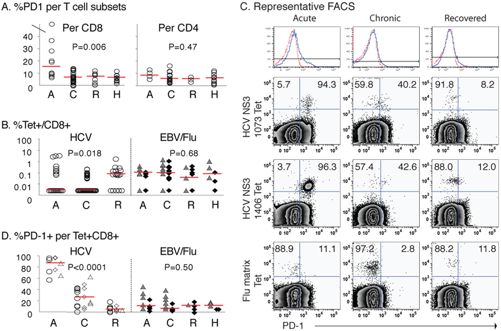
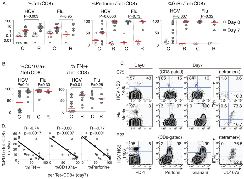
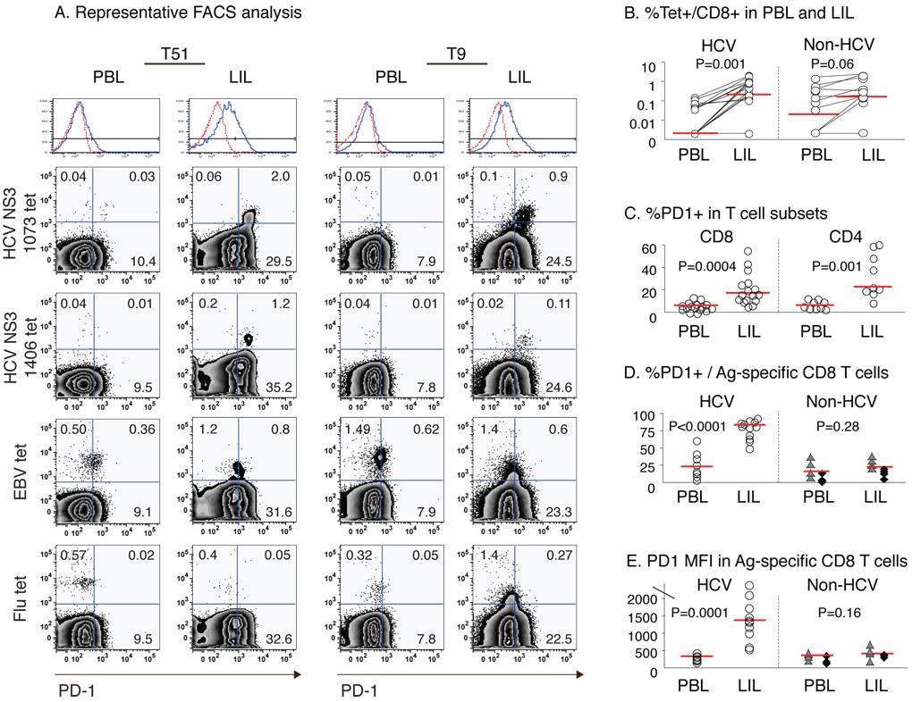
Methods: PD-1 expression and function of circulating CD8 T cells specific for HCV, Epstein-Barr virus, and influenza virus were examined ex vivo and following antigenic stimulation in vitro in patients with acute, chronic, and resolved HCV infection using class I tetramers and flow cytometry. Intrahepatic CD8 T cells were examined from liver explants of chronically HCV-infected transplant recipients.

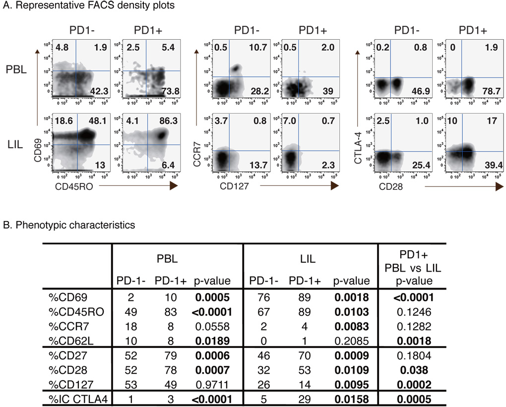
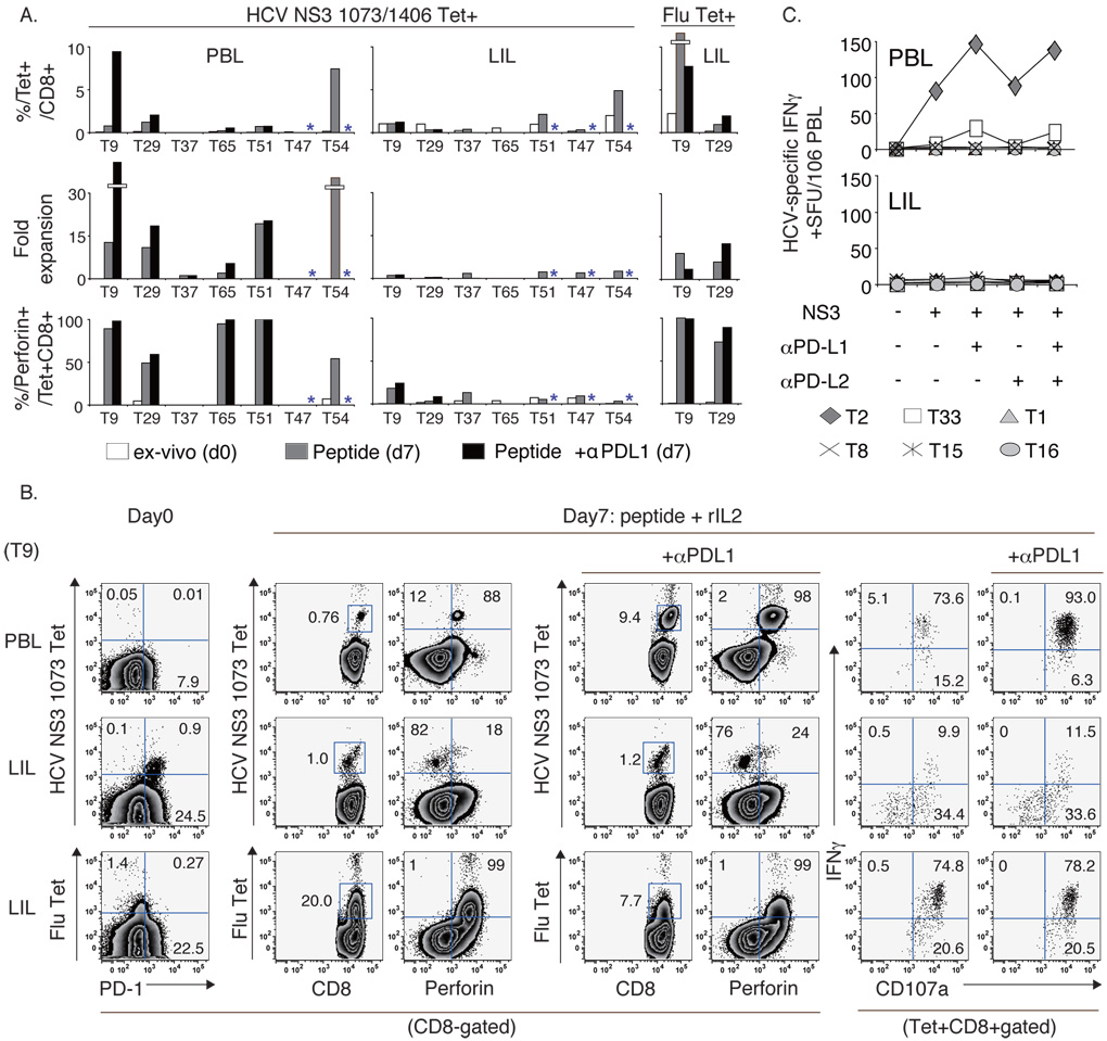
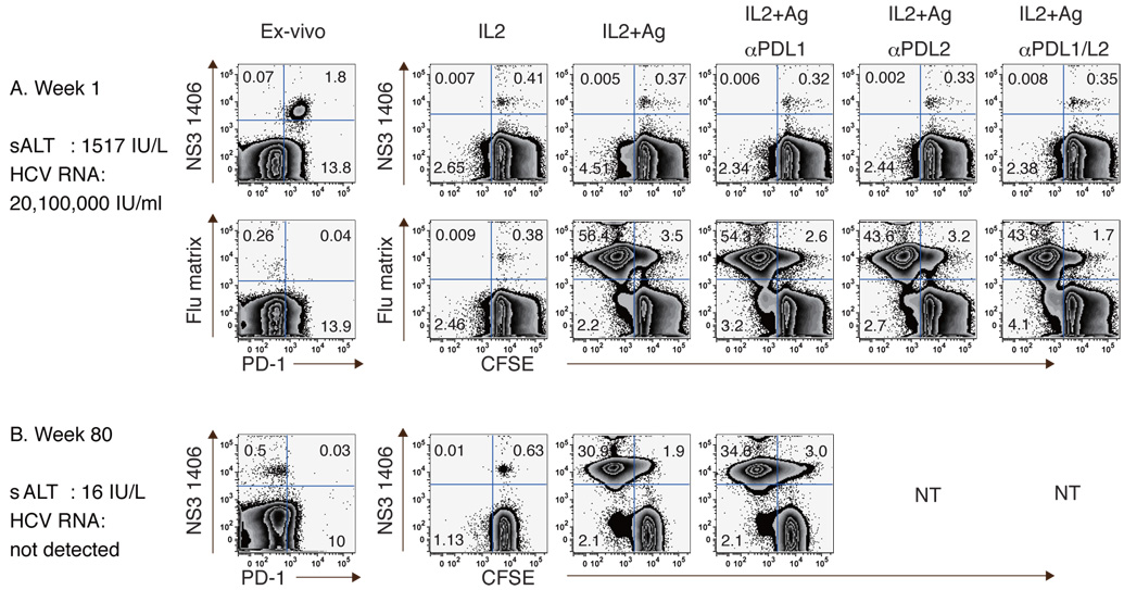
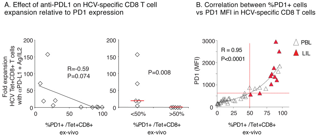
Results: Intrahepatic HCV-specific CD8 T cells from chronically HCV-infected patients were highly PD-1 positive, profoundly dysfunctional, and unexpectedly refractory to PD-1/PD-L blockade, contrasting from circulating PD-1-intermediate HCV-specific CD8 T cells with responsiveness to PD-1/PD-L blockade. This intrahepatic functional impairment was HCV-specific and directly associated with the level of PD-1 expression. Highly PD-1-positive intrahepatic CD8 T cells were more phenotypically exhausted with increased cytotoxic T-lymphocyte antigen 4 and reduced CD28 and CD127 expression, suggesting that active antigen-specific stimulation in the liver induces a profound functional exhaustion not reversible by PD-1/PD-L blockade alone.

Conclusions: HCV-specific CD8 T-cell dysfunction and responsiveness to PD-1/PD-L blockade are defined by their PD-1 expression and compartmentalization. These findings provide new and clinically relevant insight to differential antigen-specific CD8 T-cell exhaustion and their functional restoration.


Figures

Figure 1
Figure 1. Increased PD-1 expression in circulating HCV-specific CD8 T-cells from viremic patients with acute and chronic but not resolved hepatitis C
(A) %PD-1 expression in CD8 and CD4 T-cells in 10 acute (A), 27 chronic (C) and 8 resolved (R) patients with HCV infection, and 12 healthy HCV-seronegative (H) controls. Median %PD-1+/CD8 T-cells: A15.4% vs. C6.8% vs. R7.5% vs. H6.6% (p=0.006). Median %PD-1+/CD4 T-cells: A8.5% vs. C5.8% vs. R5.7% vs. H6.4% (p=0.47). (B) %Tetramer+ CD8 T-cells specific for HCV (circle), EBV (triangle), and Flu (diamond) in 7 Acute, 19 Chronic, 8 Recovered and 3 Healthy patients. Median %HCV-specific (A0.00% vs. C0.00% vs. R0.08%, p=0.018); Median %EBV or Flu-specific (A0.11% vs. C0.10% vs. R0.04% vs. H0.08, p=0.68). (C) Representative PD-1 stainings for peripheral HCV- and Flu-specific tetramer+ CD8 T-cells from acute, chronic and recovered patients. Top panel shows the PD-1 cutoff strategy with isotype control (dotted red line). (D) %PD-1+ per tetramer+ CD8 T-cells (circle, NS3 1073; diamond, NS3 1406; triangle, NS5B 2594), EBV (filled triangle) and Flu (filled diamond) in 7 acute, 19 chronic, 8 resolved and 3 healthy individuals. Median %PD-1+: HCV-specific CD8 T-cells (A87.4% vs. C26.7% vs. R5.1%, p<0.0001); non-HCV-specific CD8 T-cells (A11.1% vs. C6.5% vs. R11.8% vs. H11.8%, p=0.50). Red horizontal bars indicate the medians. P-values were determined by the Kruskal-Wallis test.
Figure 2
Figure 2. HCV-specific CD8 T-cells in patients with chronic HCV infection display impaired antigen-specific expansion and effector function in vitro
(A) HCV- and Flu-specific tetramer+ CD8 T-cell frequency and expression of perforin and granzyme B on day 0 (empty circle) and day 7 of culture (filled circle) with peptides (10µg/ml) and 100IU/ml rIL2 for 15 chronic (C) and 6 recovered (R) patients. %Tet+CD8+ (median C0.69% vs. R4.16% on day 7; p=0.003). %Perforin+/Tet+CD8+ (median C49.1% vs R92.3% on day 7, p=0.0006). %Granzyme B+/Tet+CD8+ (median C55.2% vs. R96.2% on day 7; p=0.007). Red horizontal bars indicate the median. (B) %CD107a+ and %IFNγ+ in HCV- and Flu-specific CD8 T-cells on day 7. HCV-specific CD8 T-cells: median %CD107a+ (C40.7% vs. R89.8%, p=0.01); median %IFNγ+ (C14.7% vs. R74.7%, p=0.01). (C) Representative FACS plots comparing HCV-specific and Flu-specific tetramer+ CD8 T cell expansion and effector function in chronic (C75) and resolved (R23) patients on day 0 and after 7 days of antigenic stimulation. Events are gated on CD8+ cells except for the far right intracellular staining gating on tetramer+ CD8 T-cells (D) Inverse correlation between %PD-1 expression ex vivo and antigen-specific IFNγ, CD107a and perforin expression on day 7 by HCV-specific CD8 T-cells. P-values were determined by Mann-Whitney U or Spearman Rank Correlation test).
Figure 3
Figure 3. PD-1 expression is increased in HCV-specific CD8 T-cells in the liver compared to peripheral blood
(A) Representative FACS plots for PBL and LIL from HLA-A2+ HCV-infected liver transplant recipients (T51 and T9) gating on CD8 T-cells. The top histogram shows the PD-1 cutoff strategy with isotype control (dotted red line). (B) Frequency of tetramer+CD8 T-cells specific for HCV (NS3 1073, NS3 1406, NS5B 2594), EBV and Flu epitopes in 8 HLA A2+ patients. Median values are indicated by red horizontal line. Median %HCV-specific CD8 T-cells: PBL 0.00% vs LIL 0.21%, p=0.001. Median %Flu or EBV-specific CD8 T-cells: PBL 0.02% vs. LIL 0.16%, p=0.06. (C) %PD-1 expression in CD8 and CD4 T-cells in 16 HCV-infected patients: Median %PD-1+/CD8 (PBL 6.1% vs. LIL 17.1%; p=0.0004); Median %PD-1+/CD4 (PBL 5.4% vs. LIL 21.9%; p=0.001). (D) %PD-1 expression and (E) PD-1 MFI on CD8 T-cells specific for HCV and non-HCV epitopes (EBV: triangle; Flu: diamond) from 8 HLA-A2+ HCV-infected patients. HCV-specific CD8 T cells: median %PD-1+ (PBL 23.0% vs. LIL 83.3%, p<0.0001); median PD-1 MFI (PBL 281 vs. LIL 1375, p=0.0001). Non-HCV-specific CD8 T-cells: median %PD-1+ (PBL 16.1% vs LIL 22.5%, p=0.28); median PD-1 MFI (PBL 356 vs LIL 406, p=0.16). P-values were determined by paired t-test or Mann-Whitney U.
Figure 4
Figure 4. Phenotypic characteristics of PD-1+ CD8 T-cells in peripheral blood and liver of patients with chronic HCV infection
(A) Representative FACS density plots comparing phenotypic markers for PD-1- and PD-1+ CD8 T-cells in PBL and LIL from an HCV-infected patient. Numbers in each quadrant reflect percentage of gated PD-1- and PD-1+ CD8 T-cells. (B) Phenotype of PD-1+ and PD-1− CD8 T-cells in PBL (n=24) and LIL (n=14) shown as median percentages. P-values were calculated by Mann-Whitney U.
Figure 5
Figure 5. Impaired expansion and effector function unresponsive to PD1:PD-L blockade in highly PD1+ HCV-specific CD8 T-cells in the liver of HCV-infected patients
(A) The frequency, fold expansion and perforin expression of HCV-specific and Flu-specific tetramer+ CD8 T-cells on day 0 (white bars) and on day 7 following antigenic stimulation without (gray bars) or with anti-PD-L1 (black bars) *concurrent blockade not done. All subjects were studied with NS3 1073 tetramer except T47 and T51 who displayed a dominant NS3 1406 response. (B) FACS plots showing HCV-specific and Flu-specific expansion and effector function in PBL and LIL from patient T9. (C) HCV-specific T-cell IFNγ response by IFNγ ELISpot in 6 HLA-A2-negative HCV-infected patients following stimulation with overlapping NS3 peptides +/− PD-1:PD-L blockade.
Figure 6
Figure 6. PD-1:PD-L blockade does not enhance expansion of highly PD-1+ HCV-specific CD8 T-cells in acute HCV infection
Expansion of HCV- and Flu-specific CD8 T-cells in an acute hepatitis C patient during (A) the acute (week 1) and (B) resolved phase (week 80). CFSE-labeled PBMCs were stimulated with peptides (10µg/ml) and rIL2 (100IU/ml) with/without anti-PD-L1 and/or anti-PD-L2 for 7 days.
Figure 7
Figure 7. Inverse relationship between PD-1 expression and HCV-specific CD8 T-cell expansion with anti-PD-L1 blockade
(A) Comparison of HCV tetramer+ CD8 T-cell expansion in vitro after 7 days of antigenic stimulation with anti-PD-L1 and ex vivo PD-1 expression directly (left) and in subgroups by %PD-1 cutoff of 50% (right). (B) Correlation between the frequency and MFI for PD-1 expression on HCV-specific CD8 T-cells with an exponential trendline (32 PBL, unfilled triangles; 10 LIL, red filled triangles).

Comment in

References

    1. Greenwald RJ, Freeman GJ, Sharpe AH. The B7 family revisited. Annu Rev Immunol. 2005;23:515–548. - PubMed
    1. Freeman GJ, Long AJ, Iwai Y, et al. Engagement of the PD-1 immunoinhibitory receptor by a novel B7 family member leads to negative regulation of lymphocyte activation. J Exp Med. 2000;192:1027–1034. - PMC - PubMed
    1. Freeman GJ, Wherry EJ, Ahmed R, et al. Reinvigorating exhausted HIV-specific T cells via PD-1-PD-1 ligand blockade. J Exp Med. 2006;203:2223–2227. - PMC - PubMed
    1. Barber DL, Wherry EJ, Masopust D, et al. Restoring function in exhausted CD8 T cells during chronic viral infection. Nature. 2006;439:682–687. - PubMed
    1. Day CL, Kaufmann DE, Kiepiela P, et al. PD-1 expression on HIV-specific T cells is associated with T-cell exhaustion and disease progression. Nature. 2006;443:350–354. - PubMed

Publication types

MeSH terms