. 2008 Jun;143(6):803-12.
doi: 10.1016/j.surg.2008.02.005. Epub 2008 Apr 18.

RNA stability regulates differential expression of the metastasis protein, osteopontin, in hepatocellular cancer

Affiliations

RNA stability regulates differential expression of the metastasis protein, osteopontin, in hepatocellular cancer

Sirisha Emani et al. Surgery. 2008 Jun.

Abstract

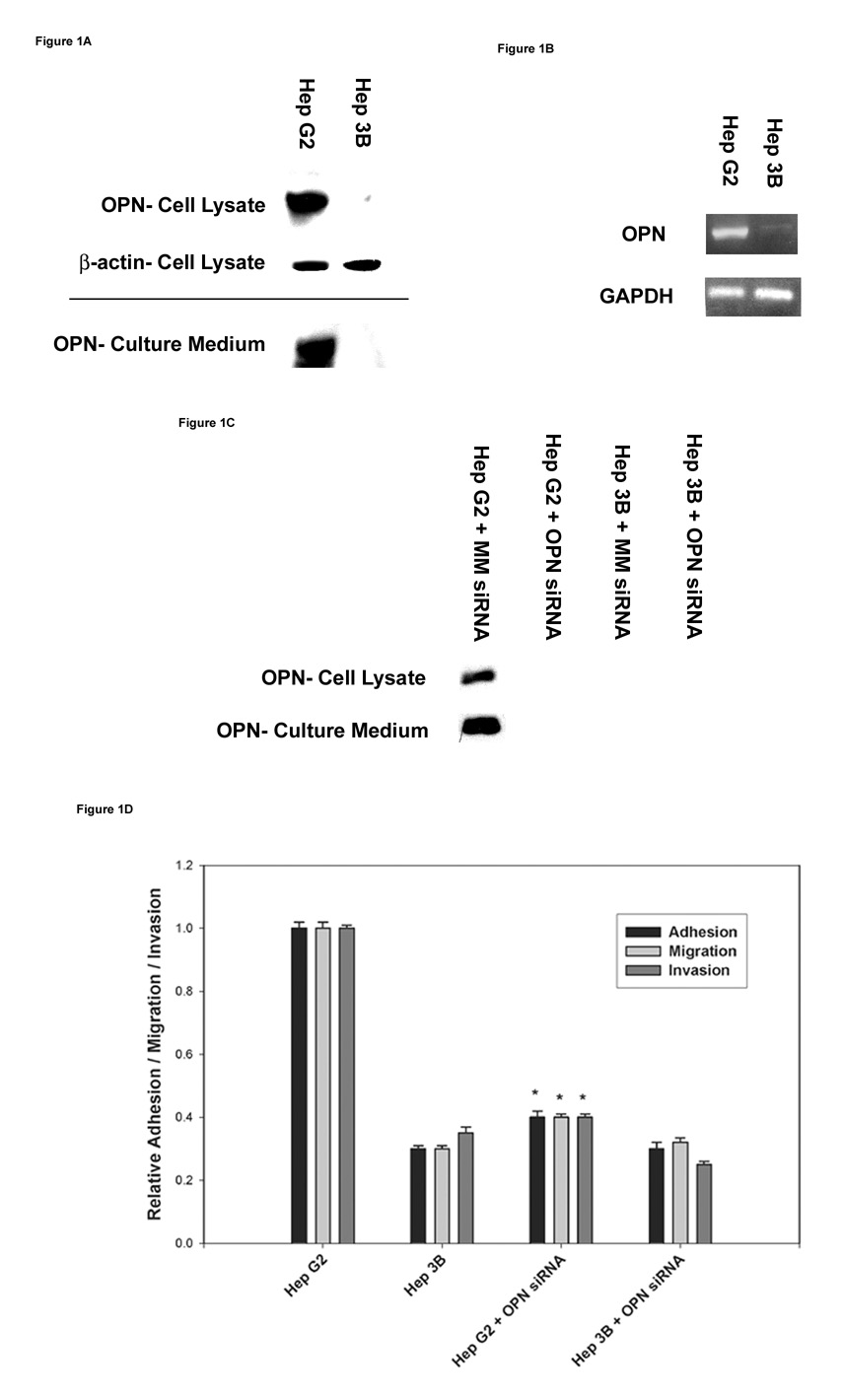
Background: Osteopontin (OPN) is a potential therapeutic target in hepatocellular carcinoma (HCC), because it is a critical mediator of metastatic function. The molecular mechanisms that determine expression of OPN in HCC, however, are unknown. In this study, we examine differential OPN expression in the 2 HCC cell lines: HepG2 and Hep3B.

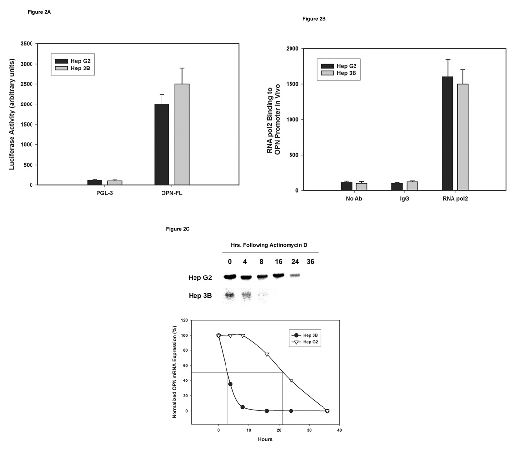
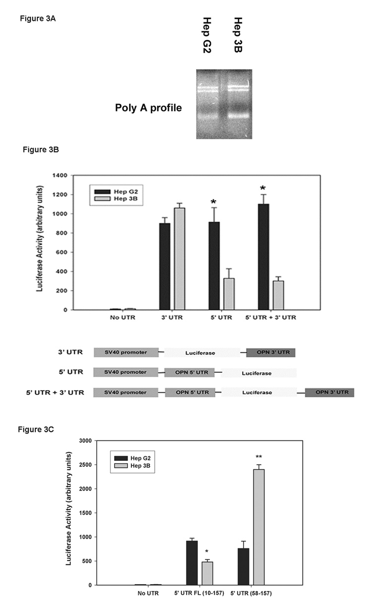
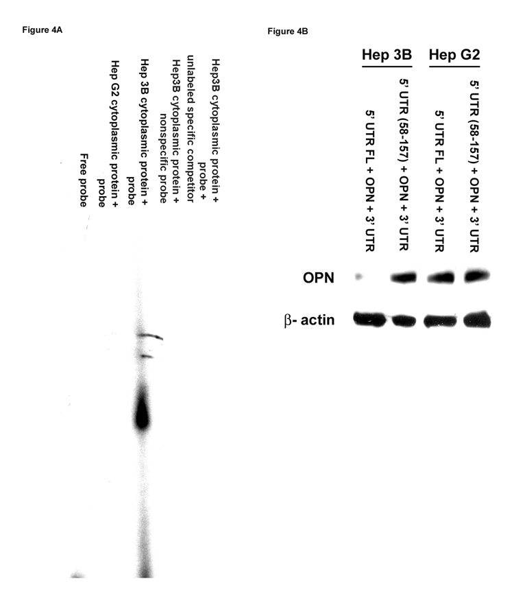
Methods: OPN expression, metastatic function, OPN promoter activity, and active transcription of OPN mRNA and its decay were assessed in the 2 HCC cell lines using standard techniques. RNA gel-shift assays were performed to determine binding of cytoplasmic proteins to OPN mRNA.

Results: Expression of OPN cellular/secreted protein and mRNA was greater in HepG2 than Hep3B cells (P < .01). Transient transfection of the OPN promoter construct demonstrated equivalent luciferase activities in the 2 cell lines; the rate of transcription was also equivalent as determined by chromatin immuno-precipitation assay. OPN mRNA half-life was 21 +/- 1 h and 3 +/- 1 h in HepG2 and Hep3B, respectively (P < .02). In HepG2 and Hep3B, the nucleotide sequence of OPN and its 5'-UTR, 3'-UTR, and poly (A) tail lengths were identical. A luciferase construct coupled in line with OPN-5'-UTR and OPN 3'-UTR presented greater expression in HepG2 (P < .01 vs Hep3B). Deletion of nt 10-57 of the OPN 5'-UTR restored luciferase and HA-tagged OPN protein expression in Hep3B but not in Hep G2. RNA gel-shift assays demonstrate different patterns of protein binding to OPN 5'-UTR between the 2 HCC cell lines.

Conclusions: We conclude that RNA stability is a new, previously unrecognized mechanism that regulates OPN expression in HCC to convey metastatic function.


Figures

Figure 1
Figure 1. Detection of OPN mRNA and protein expression in the two HCC cell lines
(A) Total cellular lysates and concentrated culture media from HepG2 and Hep3B cell lines were subjected to immunoblot analysis to determine OPN protein expression. (B) RT-PCR analysis of OPN mRNA expression in HepG2 and Hep3B cell lines. GAPDH mRNA expression was used as a house keeping control gene. (C) In vitro suppression of OPN expression by OPN siRNA transfection in the two HCC cell lines. HepG2 and Hep3B cell lines were transiently transfected with either MM siRNA (Mismatch siRNA) or OPN siRNA for 48 h. OPN protein expression was determined by western blot analysis. Blots (A–C) are representative of three separate experiments. (D) Functional properties of the two HCC cell lines were assessed by their adhesion, migration and invasion characteristics as described in materials and methods. Correlation of OPN expression to its metastatic behavior in HepG2 and Hep3B cells was determined by specifically inhibiting OPN expression by standard OPN siRNA techniques. Data are expressed as mean ± SEM (* p< 0.05 vs. HepG2).
Figure 2
Figure 2. Determination of transcription activity and OPN mRNA half-life in the two HCC cell lines
(A) HepG2 and Hep3B cells were transfected with firefly luciferase reporter plasmid vector PGL3 or PGL3 plus OPN full-length region along with 50ng/ml SV-40 expressing renilla luciferase for 48 h. Firefly luciferase activity was standardized against Renilla luciferase activity and was expressed as the mean fold luminescence for three independent experiments. (B) Binding of RNA pol2 to the OPN promoter region was assessed using ChIP assays in HepG2 and Hep3B cells followed by quantification using real-time PCR analysis; data are expressed as mean ± SEM of three independent experiments. (C) OPN mRNA half-life was determined in the two HCC cell lines in the presence of actinomycin D (20µg/ml) by Northern blot analysis after 0, 4, 8, 16, 24, and 36 h of incubation. Bands on the blots were quantified and presented in a graphical form. Blot is representative of three experiments. Graph combines data from the three experiments.
Figure 3
Figure 3. Assessment of post-transcription modifications in the OPN 5’-UTR and 3’-UTR in HepG2 and Hep3B cells
(A) Poly (A) tail profile of HepG2 and Hep3B OPN mRNA was determined by using oligo (dT) linker and T4 ligase followed by RT-PCR. PCR products were resolved on 1.2% agarose gel and visualized using ethidium-bromide staining and detection. (B) Schematic representation of OPN 3’-UTR and/or 5’-UTR cloned into luciferase containing PGL-3 plasmid vector with SV-40 promoter. Luciferase activity was determined in HepG2 and Hep3B cells transiently transfected with the above constructs (n=3). Data are expressed as mean ± SEM. (* p<0.05 vs. Hep3B). (C) Luciferase activity was performed on the HCC cell lines transiently transfected with full-length 5’-UTR as shown in Figure 3B or deletion of nt 10–57 fragment in the 5’-UTR (58–157) construct (n=3). Data are expressed as mean +/− SEM. *p values <0.05 were considered significant.
Figure 4
Figure 4. Determination of protein(s) binding to the 5’-UTR of the OPN promoter region using gel-shift assays
(A) Cytoplasmic protein lysates were extracted from HepG2 and Hep3B cells and incubated with (α-32 P) ATP (2500 Ci/mmol) end-labeled synthesized 5’-UTR from the human OPN promoter sequence using T4 polynucleotide kinase followed by G-50 column purification. The final products were resolved on 6% poly-acrylamide gel electrophoresis using 0.5× Tris/borate EDTA buffer and visualized by autoradiography. Gel is representative of three experiments. (B) Full-length OPN 5’-UTR or deletion in OPN 5’-UTR (58–157) along with OPN promoter region, OPN coding region and 3’-UTR were ligated to the pcDNA3.1 expression vector at the C-terminal followed by transient transfection of HepG2 and Hep3B cells and assayed for OPN protein expression by Western blot analysis. Blot is representative of three experiments.

Similar articles

Cited by

References

    1. Jemal A, Siegel R, Ward E, Murray T, Xu J, Thun MJ. Cancer statistics, 2007. CA Cancer J Clin. 2007;57:43–66. - PubMed
    1. Rittling SR, Chambers AF. Role of osteopontin in tumour progression. Br J Cancer. 2004;90:1877–1881. - PMC - PubMed
    1. Weber GF. The metastasis gene osteopontin: a candidate target for cancer therapy. Biochim Biophys Acta. 2001;1552:61–85. - PubMed
    1. Coppola D, Szabo M, Boulware D, Muraca P, Alsarraj M, Chambers AF, et al. Correlation of osteopontin protein expression and pathological stage across a wide variety of tumor histologies. Clin Cancer Res. 2004;10:184–190. - PubMed
    1. Chambers AF, Wilson SM, Kerkvliet N, O'Malley FP, Harris JF, Casson AG. Osteopontin expression in lung cancer. Lung cancer (Amsterdam, Netherlands) 1996;15:311–323. - PubMed

Publication types

MeSH terms