

Detailed close-ups and the big picture of spliceosomes
- PMID: 18550358
- PMCID: PMC2474778
- DOI: 10.1016/j.sbi.2008.05.005
Detailed close-ups and the big picture of spliceosomes
Abstract
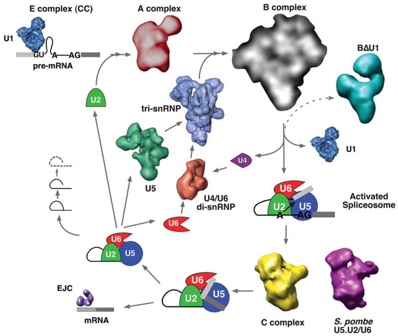
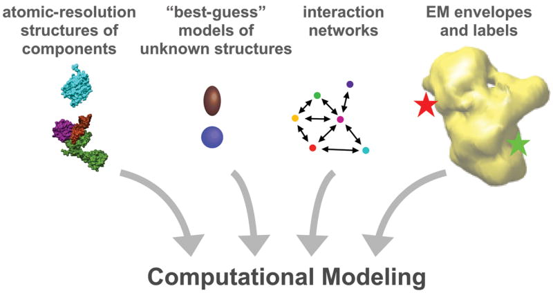
The spliceosome is the huge macromolecular assembly responsible for the removal of introns from pre-mRNA transcripts. The size and complexity of this dynamic cellular machine dictate that structural analysis of the spliceosome is best served by a combination of techniques. Electron microscopy is providing a more global, albeit less detailed, view of spliceosome assemblies. X-ray crystallographers and NMR spectroscopists are steadily reporting more atomic resolution structures of individual spliceosome components and fragments. Increasingly, structures of these individual pieces in complex with binding partners are yielding insights into the interfaces that hold the entire spliceosome assembly together. Although the information arising from the various structural studies of splicing machinery has not yet fully converged into a complete model, we can expect that a detailed understanding of spliceosome structure will arise at the juncture of structural and computational modeling methods.
Figures
References
-
- Nilsen TW. The spliceosome: the most complex macromolecular machine in the cell? Bioessays. 2003;25:1147–1149. - PubMed
-
- Moore MJ, Query CC, Sharp PA. Splicing of precursors to mRNA by the spliceosome. In: Gesteland R, Atkins J, editors. The RNA World. Cold Spring Harbor Laboratory Press; 1993. pp. 303–357.
-
- Brow DA. Allosteric cascade of spliceosome activation. Annu Rev Genet. 2002;36:333–360. - PubMed
-
- Stark H, Luhrmann R. Cryo-electron microscopy of spliceosomal components. Annu Rev Biophys Biomol Struct. 2006;35:435–457. - PubMed
Publication types
MeSH terms
Grants and funding
LinkOut - more resources
Full Text Sources
