. 2008 Aug 29;283(35):24136-44.
doi: 10.1074/jbc.M710280200. Epub 2008 Jun 11.

TRPA1 mediates the noxious effects of natural sesquiterpene deterrents

Affiliations

TRPA1 mediates the noxious effects of natural sesquiterpene deterrents

Jasmine Escalera et al. J Biol Chem. .

Abstract

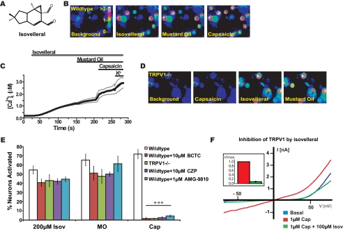
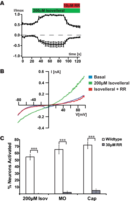
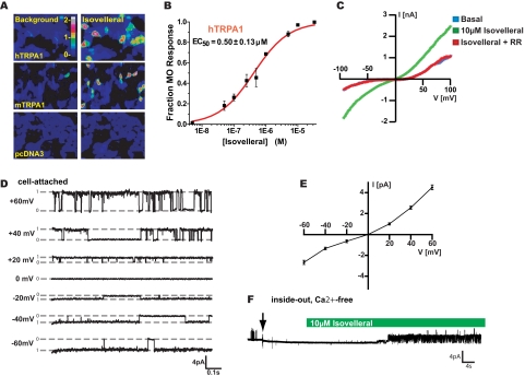
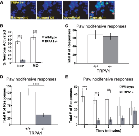
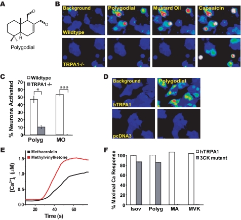
Plants, fungi, and animals generate a diverse array of deterrent natural products that induce avoidance behavior in biological adversaries. The largest known chemical family of deterrents are terpenes characterized by reactive alpha,beta-unsaturated dialdehyde moieties, including the drimane sesquiterpenes and other terpene species. Deterrent sesquiterpenes are potent activators of mammalian peripheral chemosensory neurons, causing pain and neurogenic inflammation. Despite their wide-spread synthesis and medicinal use as desensitizing analgesics, their molecular targets remain unknown. Here we show that isovelleral, a noxious fungal sesquiterpene, excites sensory neurons through activation of TPRA1, an ion channel involved in inflammatory pain signaling. TRPA1 is also activated by polygodial, a drimane sesquiterpene synthesized by plants and animals. TRPA1-deficient mice show greatly reduced nocifensive behavior in response to isovelleral, indicating that TRPA1 is the major receptor for deterrent sesquiterpenes in vivo. Isovelleral and polygodial represent the first fungal and animal small molecule agonists of nociceptive transient receptor potential channels.


Figures

FIGURE 1.
FIGURE 1.
Isovelleral-activated Ca2+ influx into cultured sensory neurons. A, structure of the pungent sesquiterpene isovelleral, the chemodefensive product of the fungus L. vellereus. B, activation of Ca2+ influx into cultured murine DRG neurons by 200 μm isovelleral as measured by fluorescent Fura-2 imaging. [Ca2+]i is represented by pseudocolors before (first panel) and 125 s after challenge with 200 μm isovelleral (second panel), followed by 100 μm mustard oil (after 50 s; third panel), and 1 μm capsaicin (after 50 s; fourth panel) at ×10 magnification. Scale bar, intracellular calcium concentration (μm). C, activation of Ca2+ influx by isovelleral into DRG neurons, plotted against time. The thick line represents the averaged [Ca2+]i in response to application of 200 μm isovelleral, followed by 100 μm mustard oil, 1 μm capsaicin, and 65 mm KCl. The thin lines represent ±S.E. Neurons (n = 342) were analyzed from three mice at ×10 magnification. D, responses of cultured DRG neurons dissociated from TRPV1-/- mice following application of 1 μm capsaicin (50 s; second panel), followed by 200 μm isovelleral (after 100 s; third panel) and 100 μm mustard oil (50 s; fourth panel). Isovelleral-induced calcium influx is retained in TRPV1-deficient sensory neurons. E, insensitivity of neuronal isovelleral-activated Ca2+ influx to pharmacological inhibition or genetic ablation of TRPV1. The bar graph shows percentages of activated neurons, in response to 200 μm isovelleral (Isov), 100 μm mustard oil (MO), and 1 μm capsaicin (Cap). The activity of agonists was tested in wild-type neurons without inhibitor (white), in the presence of the TRPV1 antagonist BCTC (10 μm; red), in TRPV1-/- neurons without inhibitor (green), and in wild-type neurons in the presence of the TRPV1 antagonists capsazepine (CZP (10 μm); purple) and AMG-9810 (1 μm; cyan). A neuron was considered to be activated when [Ca2+]i exceeded 500 nm. Values denote percentages of KCl-sensitive cells. Whereas capsaicin responsiveness was significantly reduced by antagonist and genetic ablation of TRPV1 (***, p < 0.001), isovelleral and mustard oil responsiveness remained unaffected. F, inhibition of capsaicin-activated TRPV1 currents by isovelleral. Currents from representative TRPV1-transfected HEK293t cells are shown before (blue) and after saturation of currents with 1 μm capsaicin (red). Capsaicin-activated currents were inhibited by co-application of 100 μm isovelleral (green; co-applied with capsaicin for 20 s). Currents were recorded in response to a voltage ramp (-100 mV to +100 mV for 100 ms). Inset, inward TRPV1 currents at -80 mV recorded from transfected HEK293t cells (n = 7) in the presence of capsaicin (1 μm; black) and capsaicin plus isovelleral (1 μm + 100 μm; black). Currents were normalized to the maximal capsaicin-activated current and averaged.
FIGURE 2.
FIGURE 2.
Activation of ruthenium red-sensitive ionic currents in sensory neurons by isovelleral. A, activation of ionic currents in sensory neurons by isovelleral, as measured by whole cell patch clamp electrophysiology. Currents were measured in response to a 100-ms voltage ramp from -100 to +100 mV. Averaged normalized outward and inward currents at +60 and -60 mV, respectively, of DRG neurons (n = 3) are shown measured. 200 μm isovelleral was superfused after 20 s (green bar), and 10 μm ruthenium red (RR) after 90 s (red bar). B, representative isovelleral-activated currents in a DRG neuron. The current was measured in response to a 100-ms voltage ramp from -100 to +100 mV. Blue trace, basal current before isovelleral application; green trace, fully activated currents after 200 μm isovelleral application; red trace, ruthenium red (10 μm) block of isovelleral (200 μm)-activated current. C, percentages of DRG neurons activated by 200 μm isovelleral, 100 μm mustard oil, 1 μm capsaicin, and 65 mm KCl in the presence of 30 μm ruthenium red, a TRP channel blocker (n = 147 cells with ruthenium red; n = 342 cells without ruthenium red). A neuron was considered to be activated when [Ca2+]i exceeded 500 nm.
FIGURE 3.
FIGURE 3.
Isovelleral activates TRPA1 channels in heterologous cells. A, isovelleral (10 μm) evokes calcium influx into hTRPA1- and mTRPA1 (30 μm isovelleral)-transfected cells, as measured by fluorescent Ca2+ imaging. Mock-transfected (pcDNA3) cells were insensitive to 30 μm isovelleral. Pseudocolors indicate [Ca2+]i in Fura-2-loaded cells. B, dose-response curve for activation of hTRPA1 by isovelleral in HEK293T cells, as measured by fluorescent Ca2+ imaging. Cells were activated with the indicated concentrations of isovelleral and then with a saturating dose of mustard oil (100 μm) (n = 45 ± 8 cells/dose). Error bars, S.E. C, representative whole cell currents recorded from a hTRPA1-transfected CHO cell. The current was measured in response to a 100-ms voltage ramp from -100 to +100 mV. Blue trace, basal current before isovelleral application; green trace, fully isovelleral (10 μm)-activated current, recorded 10 s following application of isovelleral to the bath solution; red trace, block of isovelleral (10 μm)-activated current by concomitant application of ruthenium red (10 μm). D, single channel currents recorded from a hTRPA1-transfected CHO cell in cell-attached configuration in response to 10 μm isovelleral. Currents were recorded from -60 to +60 mV in 20-mV steps. Average single channel conductances were 75 ± 1 picosiemens at +60 mV and 45 ± 1 picosiemens at -60 mV. E, open channel current-voltage relationship of isovelleral (10 μm)-activated hTRPA1 channels, recorded in cell-attached configuration as in D. Open channels show outward rectification. F, TRPA1 activation by isovelleral in the inside-out patch clamp configuration in Ca2+-free conditions. An inside-out patch, held at a depolarizing voltage of +40 mV, was pulled (arrow) from a CHO cell transiently transfected with hTRPA1 and perfused with 10 μm isovelleral (green bar). Bath and pipette solution contained 0 mm Ca2+ and 10 mm EGTA. TRPA1 single channels were activated by isovelleral in the absence of Ca2+. Recordings from three additional inside out patches showed similar effects (not shown).
FIGURE 4.
FIGURE 4.
Cellular and behavioral insensitivity to isovelleral in TRPA1-/- mice. A, responses of cultured DRG neurons from TRPA1-/- mice to 100 μm mustard oil (after 50 s; second panel), 200 μm isovelleral (after 150 s; third panel), and 1 μm capsaicin (after 50 s; fourth panel) as measured by Fura-2 imaging. TRPA1-deficient neurons failed to respond to mustard oil and isovelleral. B, percentages of cultured wild-type and TRPA1-/- DRG neurons activated by 200 μm isovelleral, 100 μm mustard oil, and 1 μm capsaicin. A neuron was considered to be activated when [Ca2+]i exceeded 500 nm. Residual isovelleral responses showed small changes in [Ca2+]i and had a delayed onset when compared with isovelleral-responsive wild-type neurons. ***, p < 0.001; **, p < 0.01. C, nocifensive responses of wild-type (+/+) and TRPV1-deficient (-/-) mice to paw injections of isovelleral. Responses (licking and lifting) were measured after intraplantar injection of 25 μl of 1 mm isovelleral solution into the hind paw. Responses were measured over a period of 5 min (n = 3 mice/genotype). Responses in wild-type and TRPV1-/- mice were indistinguishable. D, diminished nocifensive responses in TRPA1-deficient mice after paw injection with isovelleral. Nocifensive responses (licking, flicking, and lifting) were measured after intraplantar injection of 25 μl of 1 mm isovelleral solution into the right hind paw of mice over a period of 5 min. ***, p < 0.001 (analysis of variance), n = 10 mice/genotype. E, mouse nocifensive responses (licking, flicking, and lifting) after injection of isovelleral, plotted for each minute of the 5-min trial period. ***, p < 0.001; **, p < 0.01; n = 10 mice/genotype. Error bars, S.E.
FIGURE 5.
FIGURE 5.
TRPA1 is a receptor for polygodial and noxious terpene oxidation products. A, structure of the pungent terpene polygodial, a drimane sesquiterpene found in water pepper and other pungent plants as well as in mollusks. B, responses of DRG neurons dissociated from wild-type mice (top row) and TRPA1-/- littermates (bottom row) to 200 μm polygodial, 100 μm mustard oil, and 1 μm capsaicin, as measured by Fura-2 imaging. TRPA1-/- neurons fail to show similar increases in [Ca2+]i after exposure to polygodial but are activated by capsaicin. C, percentages of cultured wild-type and TRPA1-/- DRG neurons activated by 200 μm polygodial (Polyg), 100 μm mustard oil, and 1 μm capsaicin. A neuron was considered to be activated when [Ca2+]i exceeded 500 nm.*, p < 0.05; ***, p < 0.001. D, polygodial (10 μm) evokes calcium influx into hTRPA1-transfected cells. Cells transfected with vector alone are insensitive to 30 μm polygodial. Pseudocolor images of Fura-2-loaded cells are shown. E, trace showing calcium responses in hTRPA1-transfected cells to 100 μm methylvinylketone (red) and 100 μm methacrolein (black) as a function of time. F, requirement of covalent agonist acceptor sites in TRPA1 for activation by 30 μm isovelleral (Isov), 30 μm polygodial (Polyg), 330 μm methacrolein (MA), and 330 μm methylvinylketone (MVK). In the 3CK mutant, the amino acid residues Cys619, Cys639, Cys663, and Lys708 have been substituted with serine residues. Mutant-expressing cells were superfused with saturating doses of the compounds for 80 s, followed by saturating doses of the TRPA1 agonists carvacrol (250 μm) or 2-aminoethoxydiphenyl borate (250 μm). TRPA1 activity was measured by Fura-2 imaging. White bars, hTRPA1-transfected cells; gray bars, cells transfected with the 3CK mutant.

References

    1. Gershenzon, J., and Dudareva, N. (2007) Nat. Chem. Biol. 3 408-414 - PubMed
    1. Langenheim, J. H. (1994) J. Chem. Ecol. 20 1223-1280 - PubMed
    1. List, P. H., and Hackenberg, H. (1969) Arch. Pharm. 302 125-143 - PubMed
    1. Magnusson, G., Thoren, S., and Wickberg, B. (1972) Tetrahedron Lett. 13 1105-1108
    1. Camazine, S. M., Resch, J. F., Eisner, T., and Meinwald, J. (1983) J. Chem. Ecol. 9 1439-1447 - PubMed

Publication types