. 2008 Aug;295(2):C414-22.
doi: 10.1152/ajpcell.00038.2008. Epub 2008 Jun 11.

Cytoplasmic cAMP concentrations in intact cardiac myocytes

Affiliations

Cytoplasmic cAMP concentrations in intact cardiac myocytes

Radu V Iancu et al. Am J Physiol Cell Physiol. 2008 Aug.

Abstract

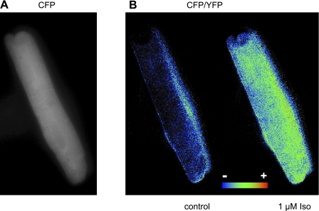
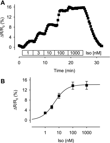
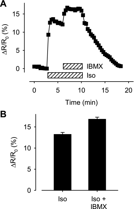
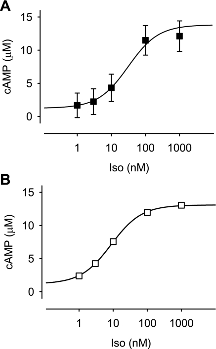
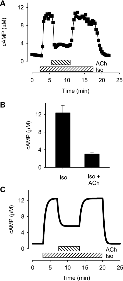
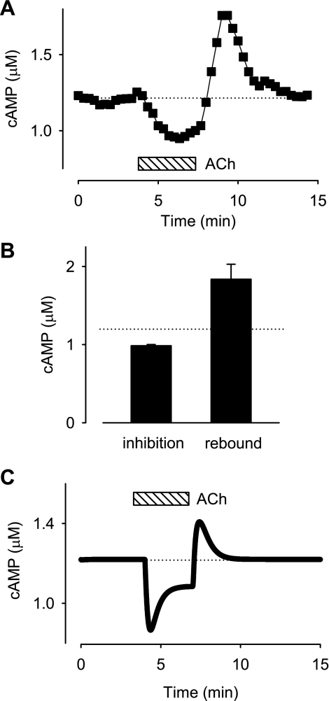
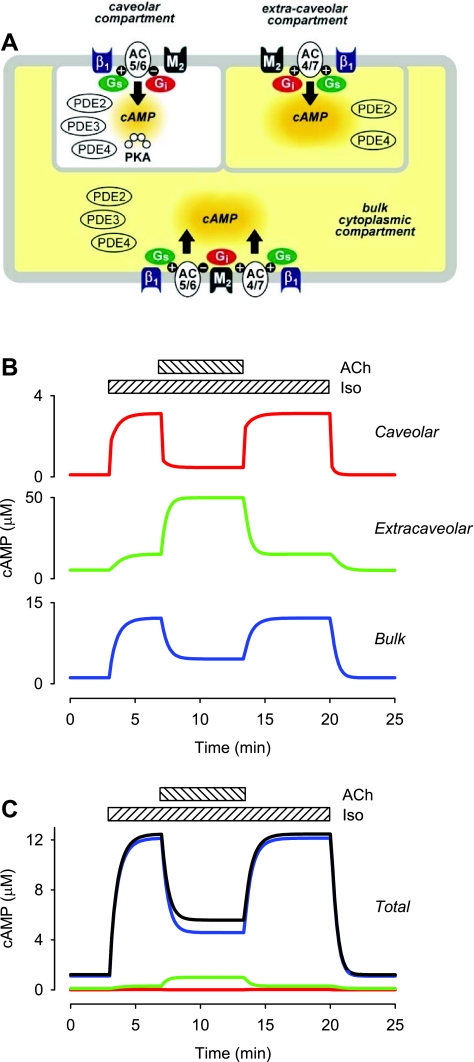
In cardiac myocytes there is evidence that activation of some receptors can regulate protein kinase A (PKA)-dependent responses by stimulating cAMP production that is limited to discrete intracellular domains. We previously developed a computational model of compartmentalized cAMP signaling to investigate the feasibility of this idea. The model was able to reproduce experimental results demonstrating that both beta(1)-adrenergic and M(2) muscarinic receptor-mediated cAMP changes occur in microdomains associated with PKA signaling. However, the model also suggested that the cAMP concentration throughout most of the cell could be significantly higher than that found in PKA-signaling domains. In the present study we tested this counterintuitive hypothesis using a freely diffusible fluorescence resonance energy transfer-based biosensor constructed from the type 2 exchange protein activated by cAMP (Epac2-camps). It was determined that in adult ventricular myocytes the basal cAMP concentration detected by the probe is approximately 1.2 muM, which is high enough to maximally activate PKA. Furthermore, the probe detected responses produced by both beta(1) and M(2) receptor activation. Modeling suggests that responses detected by Epac2-camps mainly reflect what is happening in a bulk cytosolic compartment with little contribution from microdomains where PKA signaling occurs. These results support the conclusion that even though beta(1) and M(2) receptor activation can produce global changes in cAMP, compartmentation plays an important role by maintaining microdomains where cAMP levels are significantly below that found throughout most of the cell. This allows receptor stimulation to regulate cAMP activity over concentration ranges appropriate for modulating both higher (e.g., PKA) and lower affinity (e.g., Epac) effectors.


Figures

Fig. 1.
Fig. 1.
Guinea pig ventricular myocyte 48 h after infection with adenovirus expressing the type 2 exchange protein activated by cAMP (Epac2- camps) biosensor. A: expression pattern illustrated by cyan fluorescent protein (CFP) fluorescence. B: pseudocolor image of the CFP-to yellow fluorescent protein (YFP) (CFP/YFP) fluorescence ratio before and after exposure to a maximally stimulating concentration (1 μM) of the β-adrenergic receptor agonist isoproterenol (Iso).
Fig. 2.
Fig. 2.
Concentration dependence of the type 2 exchange protein activated by cAMP (Epac2-camps) response to β-adrenergic receptor stimulation with Iso. A: time course of the relative change in CFP/YFP emission intensity ratio (ΔR/R0) produced by Epac2-camps during exposure of a ventricular myocyte to increasing concentrations of Iso. B: concentration response relationship for the Epac2-camps response to Iso. Data points were fit by a Hill equation with the following parameters: EC50, 4.7 nM; maximum response, 14%; Hill coefficient, 1.
Fig. 3.
Fig. 3.
Maximum response of Epac2-camps in intact cells. A: time course of changes in the relative CFP/YFP emission intensity ratio (ΔR/R0) produced by Epac2 sensor during exposure to a maximally stimulating concentration (1 μM) of the β-adrenergic receptor agonist Iso followed by subsequent addition of a maximally effective concentration (100 μM) of the nonselective phosphodiesterase inhibitor 3-isobutyl-1-methylxanthine (IBMX). B: average responses to 1 μM Iso and Iso plus 100 μM IBMX (P < 0.05).
Fig. 4.
Fig. 4.
Concentration of cAMP detected by Epac2-camps in adult ventricular myocytes. A: estimated concentration of cAMP produced by different concentrations of the β-adrenergic receptor agonist Iso. Relative CFP/YFP emission intensity ratio measurements from Fig. 2B were converted to cAMP concentration by Eq. 1. Parameters of fit to experimental data points: EC50, 31 nM Iso; minimum cAMP concentration, 1.2 μM; maximum cAMP concentration, 13.5 μM; Hill coefficient, 1. B: effect of Iso on the total cAMP concentration predicted by computational model of cAMP signaling in an adult ventricular myocyte (see methods). Parameters of fit to predicted data points: EC50, 8.8 nM Iso; minimum cAMP concentration, 1.2 μM; maximum cAMP concentration, 13.1 μM; and Hill coefficient 1. Note: experimental and predicted values are consistent with one another.
Fig. 5.
Fig. 5.
Muscarinic inhibition of β-adrenergic response. A: time course of changes in total cAMP concentration in a ventricular myocyte exposed to a maximally stimulating concentration (0.2 μM) of Iso and subsequent addition of 10 μM acetylcholine (ACh). The concentration of cAMP detected by Epac2-camps was estimated using Eq. 1. B: averaged response to 0.2 μM Iso and Iso plus 10 μM ACh (P < 0.05). C: effects of Iso and ACh on the total cAMP concentration predicted by computational model of cAMP signaling in an adult ventricular myocyte (see methods). Note: baseline cAMP concentration, cAMP concentration in the presence of a maximally stimulating concentration of Iso, and cAMP concentration in the presence of Iso plus ACh are consistent with experimental observations.
Fig. 6.
Fig. 6.
Muscarinic inhibition and stimulation of basal cAMP. A: time course of changes in cAMP concentration during exposure to 10 μM ACh and following ACh washout. The concentration of cAMP detected by Epac2-camps was estimated by Eq. 1. B: average inhibitory response observed in the presence of 10 μM ACh and peak rebound stimulatory response observed after washout of ACh. C: effects of ACh on basal cAMP concentration predicted by computational model (see methods). Note: steady-state inhibition of basal cAMP concentration by exposure to ACh and rebound increase in cAMP concentration following washout of ACh are consistent with experimental observations.
Fig. 7.
Fig. 7.
Computational modeling of cAMP concentrations in different microdomains in adult ventricular myocytes. A: diagram of different subcellular compartments included in computational model (relative size is not drawn to scale): β1, β1-adrenergic receptor; M2, M2 muscarinic receptor; AC5/6, adenylyl cyclase 5 and/or 6; AC4/7, adenylyl cyclase 4 and/or 7; Gi, inhibitory G protein; Gs, stimulatory G protein. See text for full details. B: effect of 0.2 μM Iso and 10 μM ACh on time course of changes in cAMP concentration in the different subcellular compartments. C: relative contribution of each subcellular compartment to total cAMP concentration.

References

    1. Adams SR, Harootunian AT, Buechler YJ, Taylor SS, Tsien RY. Fluorescence ratio imaging of cyclic AMP in single cells. Nature 349: 694–697, 1991. - PubMed
    1. Belevych AE, Juranek I, Harvey RD. Protein kinase C regulates functional coupling of β1-adrenergic receptors to Gi/o-mediated responses in cardiac myocytes. FASEB J 18: 367–369, 2004. - PubMed
    1. Belevych AE, Sims C, Harvey RD. ACh-induced rebound stimulation of L-type Ca2+ current in guinea-pig ventricular myocytes, mediated by Gbetagamma-dependent activation of adenylyl cyclase. J Physiol 536: 677–692, 2001. - PMC - PubMed
    1. Bers DM Excitation-Contraction Coupling and Cardiac Contractile Force. Dordrecht, The Netherlands: Kluwer, 2001.
    1. Bos JL EpaC proteins: multi-purpose cAMP targets. Trends Biochem Sci 31: 680–686, 2006. - PubMed

Publication types

MeSH terms

LinkOut - more resources