

Contextual fear conditioning in humans: cortical-hippocampal and amygdala contributions
- PMID: 18550763
- PMCID: PMC2475649
- DOI: 10.1523/JNEUROSCI.1246-08.2008
Contextual fear conditioning in humans: cortical-hippocampal and amygdala contributions
Abstract
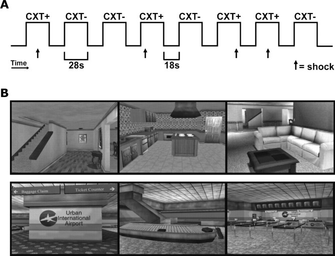
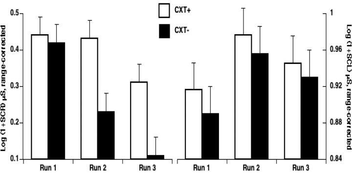
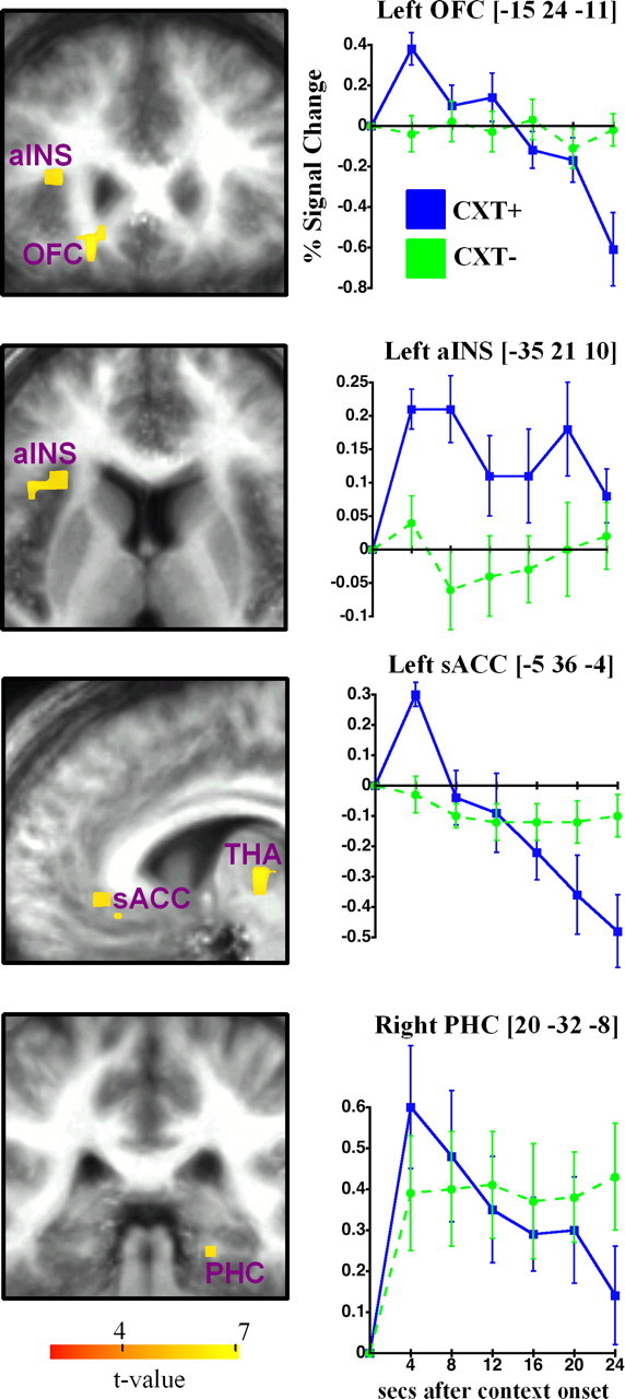
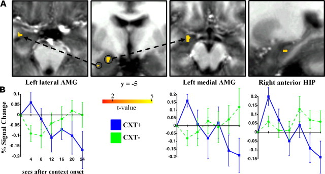
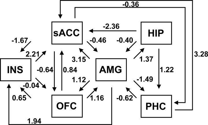
Functional imaging studies of cued fear conditioning in humans have mostly confirmed findings in animals, but it is unclear whether the brain mechanisms that underlie contextual fear conditioning in animals are also preserved in humans. We investigated this issue using functional magnetic resonance imaging and virtual reality contexts. Subjects underwent differential context conditioning in which they were repeatedly exposed to two contexts (CXT+ and CXT-) in semirandom order, with contexts counterbalanced across participants. An unsignaled footshock was consistently paired with the CXT+, and no shock was ever delivered in the CXT-. Evidence for context conditioning was established using skin conductance and anxiety ratings. Consistent with animal models centrally implicating the hippocampus and amygdala in a network supporting context conditioning, CXT+ compared with CXT- significantly activated right anterior hippocampus and bilateral amygdala. In addition, context conditioning was associated with activation in posterior orbitofrontal cortex, medial dorsal thalamus, anterior insula, subgenual anterior cingulate, and parahippocampal, inferior frontal, and parietal cortices. Structural equation modeling was used to assess interactions among the core brain regions mediating context conditioning. The derived model indicated that medial amygdala was the source of key efferent and afferent connections including input from orbitofrontal cortex. These results provide evidence that similar brain mechanisms may underlie contextual fear conditioning across species.
Figures
References
-
- Amaral DG, Price JL. Amygdalo-cortical projections in the monkey (Macaca fascicularis) J Comp Neurol. 1984;230:465–496. - PubMed
-
- Bannerman DM, Grubb M, Deacon RM, Yee BK, Feldon J, Rawlins JN. Ventral hippocampal lesions affect anxiety but not spatial learning. Behav Brain Res. 2003;139:197–213. - PubMed
-
- Barbas H, Blatt GJ. Topographically specific hippocampal projections target functionally distinct prefrontal areas in the rhesus monkey. Hippocampus. 1995;5:511–533. - PubMed
-
- Bohlin G. Delayed habituation of the electrodermal orienting response as a function of increased level of arousal. Psychophysiology. 1976;13:345–351. - PubMed
Publication types
MeSH terms
Substances
Grants and funding
LinkOut - more resources
Full Text Sources
Other Literature Sources
Medical