. 2008 Jun 17;105(24):8256-61.
doi: 10.1073/pnas.0801340105. Epub 2008 Jun 11.

Effect of glycosylation on protein folding: a close look at thermodynamic stabilization

Affiliations

Effect of glycosylation on protein folding: a close look at thermodynamic stabilization

Dalit Shental-Bechor et al. Proc Natl Acad Sci U S A. .

Abstract

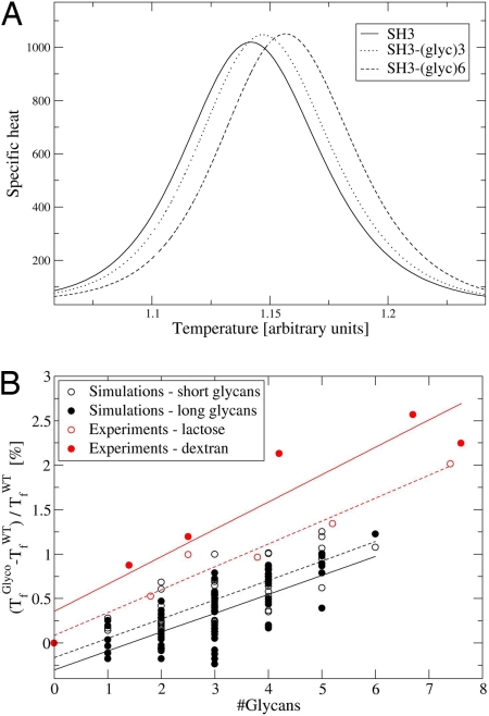
Glycosylation is one of the most common posttranslational modifications to occur in protein biosynthesis, yet its effect on the thermodynamics and kinetics of proteins is poorly understood. A minimalist model based on the native protein topology, in which each amino acid and sugar ring was represented by a single bead, was used to study the effect of glycosylation on protein folding. We studied in silico the folding of 63 engineered SH3 domain variants that had been glycosylated with different numbers of conjugated polysaccharide chains at different sites on the protein's surface. Thermal stabilization of the protein by the polysaccharide chains was observed in proportion to the number of attached chains. Consistent with recent experimental data, the degree of thermal stabilization depended on the position of the glycosylation sites, but only very weakly on the size of the glycans. A thermodynamic analysis showed that the origin of the enhanced protein stabilization by glycosylation is destabilization of the unfolded state rather than stabilization of the folded state. The higher free energy of the unfolded state is enthalpic in origin because the bulky polysaccharide chains force the unfolded ensemble to adopt more extended conformations by prohibiting formation of a residual structure. The thermodynamic stabilization induced by glycosylation is coupled with kinetic stabilization. The effects introduced by the glycans on the biophysical properties of proteins are likely to be relevant to other protein polymeric conjugate systems that regularly occur in the cell as posttranslational modifications or for biotechnological purposes.


Conflict of interest statement

The authors declare no conflict of interest.

Figures

Fig. 1.
Fig. 1.
Schematic free-energy profile of a glycoprotein. Glycosylation may result in higher stability compared with the nonglycosylated wild-type protein (i.e., ΔGGlycoGWT). The higher stability can be the outcome of a less stable unfolded state (i.e., ΔGUGlyco−ΔGUWT>0) or a more stable folded state (i.e., ΔGFGlyco−ΔGFWT<0). Glycosylation also may affect the height of the folding barrier.
Fig. 2.
Fig. 2.
Ribbon diagram of the SH3 domain with six “designed” polysaccharide chains. The polypeptide chain is colored blue, and the carbohydrate rings are represented as gray balls. Each glycan contains 11 sugar rings. In the coarse-grained model, each sugar ring is represented by a single bead with a radius of 6 Å.
Fig. 3.
Fig. 3.
Thermal stabilization of the glycosylated variants of the SH3 domain. (A) Specific heat curves of native SH3 (filled line) and of SH3 modified with three (dotted line) and six (dashed line) 11-sugar ring glycans. (B) Effect of glycosylation on protein stability as measured by the change in the Tf. Simulations of 63 SH3 glycosylated variants bearing short glycans comprised 5 sugar rings (open black circles, fitted by a dashed black line; R = 0.805) or long glycans comprised of 11 sugar rings (filled black circles, fitted by a solid black line; R = 0.719). Experimental measurements of stabilization by chemical glycosylation of α-CT (27) with lactose (open red circles, fitted by a dashed red line; R = 0.98) or 10-kDa dextran (filled red circles, fitted by a solid red line; R = 0.936) from the work of Sola et al. (27, 28) are shown. The number of glycans conjugated to α-CT was calculated as the average moles of glycan per mole of α-CT.
Fig. 4.
Fig. 4.
Glycosylation effect on the free energy of unfolding. (A) Free-energy profile against the fraction of native contacts (Q used as a reaction coordinate) for native SH3 (filled line) and for SH3 glycosylated with three (dotted line) or six (dashed line) 11-sugar ring glycans. (B) Kinetic effect of glycosylation as measured by the change in the height of the free-energy barrier of unfolding (at the Tf of the native SH3 domain). (C) Free-energy barrier of unfolding at the specific Tf of each glyco-conjugated variant of the SH3 domain (divided by the specific Tf).
Fig. 5.
Fig. 5.
Effect of glycosylation on the enthalpy (H) and entropy (TS) of the folded and unfolded states. (A) Free energy of folding ΔGf (black) of the SH3 domain and its 63 glycosylated variants as a function of the number of added glycans and the free energy of the folded (Gf, gray) and unfolded (Gun, blue) ensembles. The energetic analysis was performed at the Tf of the nonglycosylated SH3 domain. (B) Enthalpic (gray) and entropic (brown) contributions to the free energy of the folded state ensemble as a function of the number of added glycans. (C) Enthalpic (gray) and entropic (brown) contributions to the free energy of the unfolded state ensemble as a function of the number of added glycans.
Fig. 6.
Fig. 6.
Effect of glycosylation on the structure of the folded and unfolded state ensembles. (A and B) Structure of the folded (A) and unfolded (B) states measured as the difference between the number of native contacts that are formed by each specific residue of the glycan-conjugated protein and the native SH3 domain. The arrows indicate the positions of the glycosylation sites. The term piSH3 represents the probability of each residue of the nonglycosylated SH3-forming native contacts, whereas piglyco is the equivalent term for the glycosylated variants.

Similar articles

Cited by

References

    1. Fersht AR, Daggett V. Protein folding and unfolding at atomic resolution. Cell. 2002;108:573–582. - PubMed
    1. Oliveberg M, Wolynes PG. The experimental survey of protein-folding energy landscapes. Q Rev Biophys. 2005;38:245–288. - PubMed
    1. Onuchic JN, Wolynes PG. Theory of protein folding. Curr Opin Struct Biol. 2004;14:70–75. - PubMed
    1. Papoian GA, Ulander J, Eastwood MP, Luthey-Schulten Z, Wolynes PG. Water in protein structure prediction. Proc Natl Acad Sci USA. 2004;101:3352–3357. - PMC - PubMed
    1. Jin W, Kambara O, Sasakawa H, Tamura A, Takada S. De novo design of foldable proteins with smooth folding funnel: Automated negative design and experimental verification. Structure. 2003;11:581–590. - PubMed

Publication types