. 2008 Jun 17;105(24):8274-9.
doi: 10.1073/pnas.0803330105. Epub 2008 Jun 12.

Saccharomyces cerevisiae septins: supramolecular organization of heterooligomers and the mechanism of filament assembly

Affiliations

Saccharomyces cerevisiae septins: supramolecular organization of heterooligomers and the mechanism of filament assembly

Aurelie Bertin et al. Proc Natl Acad Sci U S A. .

Abstract

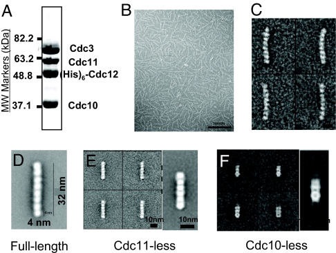
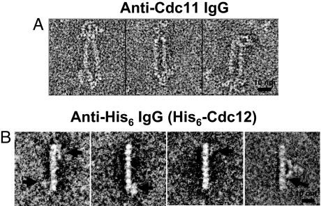
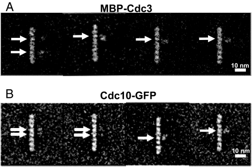
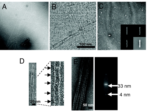
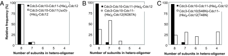
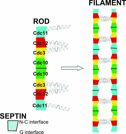
Mitotic yeast cells express five septins (Cdc3, Cdc10, Cdc11, Cdc12, and Shs1/Sep7). Only Shs1 is nonessential. The four essential septins form a complex containing two copies of each, but their arrangement was not known. Single-particle analysis by EM confirmed that the heterooligomer is octameric and revealed that the subunits are arrayed in a linear rod. Identity of each subunit was determined by examining complexes lacking a given septin, by antibody decoration, and by fusion to marker proteins (GFP or maltose binding protein). The rod has the order Cdc11-Cdc12-Cdc3-Cdc10-Cdc10-Cdc3-Cdc12-Cdc11 and, hence, lacks polarity. At low ionic strength, rods assemble end-to-end to form filaments but not when Cdc11 is absent or its N terminus is altered. Filaments invariably pair into long parallel "railroad tracks." Lateral association seems to be mediated by heterotetrameric coiled coils between the paired C-terminal extensions of Cdc3 and Cdc12 projecting orthogonally from each filament. Shs1 may be able to replace Cdc11 at the end of the rod. Our findings provide insights into the molecular mechanisms underlying the function and regulation of cellular septin structures.


Conflict of interest statement

The authors declare no conflict of interest.

Figures

Fig. 1.
Fig. 1.
Characterization of yeast heterooligomeric septin complexes. (A) A sample (50 μg) of the purified Cdc3–Cdc10–Cdc11–(His)6–Cdc12 heterooctamers used in most of these studies, prepared as described in Materials and Methods, was resolved by electrophoresis on a 10% SDS/PAGE gel and stained with Coomassie Brilliant blue. MW, molecular weight. (B) Low magnification view of the preparation shown in (A) suspended in high-salt (300 mM) buffer, adsorbed on a carbon grid, stained, and viewed in the EM as described in Materials and Methods. (C) Four representative class averages (≈150 particles each) derived from processing a total of 11,000 particles from micrographs of Cdc3–Cdc10–Cdc11–(His)6–Cdc12 complexes, as in (B). (D) Higher magnification view of an average of 1,986 particles of the Cdc3–Cdc10–Cdc11–(His)6–Cdc12 (full-length) complex. (E) Four representative class averages (100 particles each) (Left) and a higher magnification view of the average of the whole data set of 2,093 particles (Right) observed in a preparation of Cdc3–Cdc10–(His)6–Cdc12 (Cdc11-less) complexes, examined as in B. (F) Four representative class averages (10 particles each) (Left) and a higher magnification view of the average of the whole data set of 824 particles (Right) observed in a preparation of Cdc3–Cdc11–(His)6–Cdc12 (Cdc10-less) complexes, examined as in B.
Fig. 2.
Fig. 2.
Location of Cdc11 and (His)6–Cdc12 determined by antibody decoration. (A) A sample of the preparation of purified Cdc3–Cdc10–Cdc11–(His)6–Cdc12 heterooctamers shown in Fig. 1 A–D was incubated on ice in high-salt buffer with rabbit polyclonal anti-Cdc11 antibodies. Three representative raw (unaveraged) images of the resulting complexes observed are shown. (B) As in A, except incubation was with a mouse monoclonal anti-(His)6 antibody. Although binding of this antibody was substoichiometric under the high-salt conditions, image processing readily identified particles with antibody bound. Left, three representative raw (unaveraged) images of the resulting complexes observed are shown, wherein the extra density (horizontal arrows) is most often positioned closest to the second subunit in from one or both ends of the rod. Right, one of the class averages obtained after multiple rounds of multireference alignment and classification, in which the antibody is seen bound symmetrically around the center of the rod, with at least one of its Fab arms in contact with the second subunit from the end (most clearly seen for the contact indicated by the arrow).
Fig. 3.
Fig. 3.
Location of MBP–Cdc3 and Cdc10–GFP. (A) Four representative class averages of purified MBP–Cdc3–Cdc10–Cdc11–(His)6–Cdc12 heterooctamers resulting from three rounds of multireference alignment and classification (2,620 particles total). The extra density (horizontal arrows) is most often positioned closest to the third subunit in from one or both ends of the rod. (B) Four representative class averages of purified Cdc3–Cdc10–GFP–Cdc11–(His)6–Cdc12 heterooctamers resulting from three rounds of multireference alignment and classification (3,377 particles total). The extra density (horizontal arrows) is most often positioned closest to one or both of the two central subunits in the rod. In A and B, the majority of the averages yielded only one extra density per octamer, most likely because of flexibility in the joint between the marker protein and septin, making detection of the second marker improbable.
Fig. 4.
Fig. 4.
End-on-end polymerization of the rods forms paired filaments. (A) A sample of the purified Cdc3–Cdc10–Cdc11–(His)6–Cdc12 heterooctamers shown in Fig. 1 A–D were diluted from high salt (300 mM) to low salt (50 mM), incubated for 2 h, and then examined in the EM after negative staining. (B) As in A, except that the sample was viewed via cryo-EM after vitrification in liquid ethane, as described in Materials and Methods. (C) Purified Cdc3–Cdc10–Cdc11(Δα0)–(His)6–Cdc12 heterooctamers were examined after dilution and incubation in low salt, as in A. (Inset) Three representative class averages and the average of the most common class (Bottom Right) of the same sample of purified Cdc3–Cdc10–Cdc11(Δα0)–(His)6–Cdc12 heterooctamers examined in high salt. (D) Paired filaments displaying periodic densities between the filaments (Left) and at higher magnification (Right). (E) Example of a bundle of paired filaments formed after prolonged incubation under low-salt conditions at relatively high protein concentration (>1 μg ml−1) and above neutral pH (Left) and a representative power spectrum (Right) derived from the Fourier transform of the image of such a bundle of paired filaments.
Fig. 5.
Fig. 5.
Effect of site-directed mutations on the efficiency of stable heterooctameric rod assembly. Purified preparations of the wild-type Cdc3–Cdc10–Cdc11–(His)6–Cdc12 complex (black bars) and the three mutant complexes indicated in each panel (white bars), Cdc3–Cdc10–Cdc11(Δα0)–(His)6–Cdc12 (A), Cdc3–Cdc10–Cdc11–(His)6–Cdc12(W267A) (B), and Cdc3–Cdc10(S46N)–Cdc11–(His)6–Cdc12(T48N) (C), were examined under high-salt conditions in the EM after negative staining, and the number of subunits in the rods formed were scored; 3,043 particles of the Cdc3–Cdc10–Cdc11(Δα0)–(His)6–Cdc12 complexes, 2,130 total particles for the Cdc3–Cdc10–Cdc11–(His)6–Cdc12(W267A) complexes, and 1,267 particles for the Cdc3–Cdc10(S46N)–Cdc11–(His)6–Cdc12(T48N) complexes were selected, aligned, and classified, first without any reference before a second round of alignment and classification by using references resulting from the first round of alignment, and classification was performed.
Fig. 6.
Fig. 6.
Schematic diagram of the yeast septin rod, filament, and paired filament assemblies. The yeast heterooctameric septin complex is a linear rod with the subunits arrayed in the order and with the interfaces indicated. Each septin forms associations with its neighbors through either a G interface or an N–C interface. The rod is nonpolar because it has a two-fold axis of rotational symmetry running left-to-right between and orthogonal to the central pair of Cdc10 septins. The CTEs of the two Cdc3 and Cdc12 pairs project from the same face of the rod and presumably associate to form parallel coiled coils that are essential for rod stability because no rod assembly or filament formation occurs in their absence. Filaments form via end-on-end assembly of the rods mediated by an ionic strength-dependent Cdc11–Cdc11 interaction through an N–C interface. The CTE of Cdc11 is not required either for rod assembly or for filament formation. Filament formation and pairing in register are coupled, with the pairing presumably mediated by lateral association between the parallel Cdc3–Cdc12 coiled coil on one filament with a corresponding parallel Cdc3–Cdc12 coiled coil on the neighboring filament, thereby forming an antiparallel four-helix bundle.

References

    1. Pan F, et al. Analysis of septins across kingdoms reveals orthology and new motifs. BMC Evol Biol. 2007;7:103.1–103.17. - PMC - PubMed
    1. Versele M, Thorner J. Some assembly required: Yeast septins provide the instruction manual. Trends Cell Biol. 2005;15:414–424. - PMC - PubMed
    1. Hartwell LH. Genetic control of the cell division cycle in yeast. IV. Genes controlling bud emergence and cytokinesis. Exp Cell Res. 1971;69:265–276. - PubMed
    1. Harbury PB, et al. A switch between two-, three-, and four-stranded coiled coils in GCN4 leucine zipper mutants. Science. 1993;262:1401–1407. - PubMed
    1. Versele M, et al. Protein-protein interactions governing septin heteropentamer assembly and septin filament organization in Saccharomyces cerevisiae. Mol Biol Cell. 2004;15:4568–4583. - PMC - PubMed

Publication types

MeSH terms