. 2008 Jun 13;4(6):e1000084.
doi: 10.1371/journal.ppat.1000084.

The malaria secretome: from algorithms to essential function in blood stage infection

Affiliations

The malaria secretome: from algorithms to essential function in blood stage infection

Christiaan van Ooij et al. PLoS Pathog. .

Erratum in

  • PLoS Pathog. 2008 Jun;4(6). doi: 10.1371/annotation/2b000375-e083-46ed-a44a-ff297e6c37d0. Waters, Andy [corrected to Waters, Andrew P]; Janse, Chris [corrected to Janse, Chris J]

Abstract

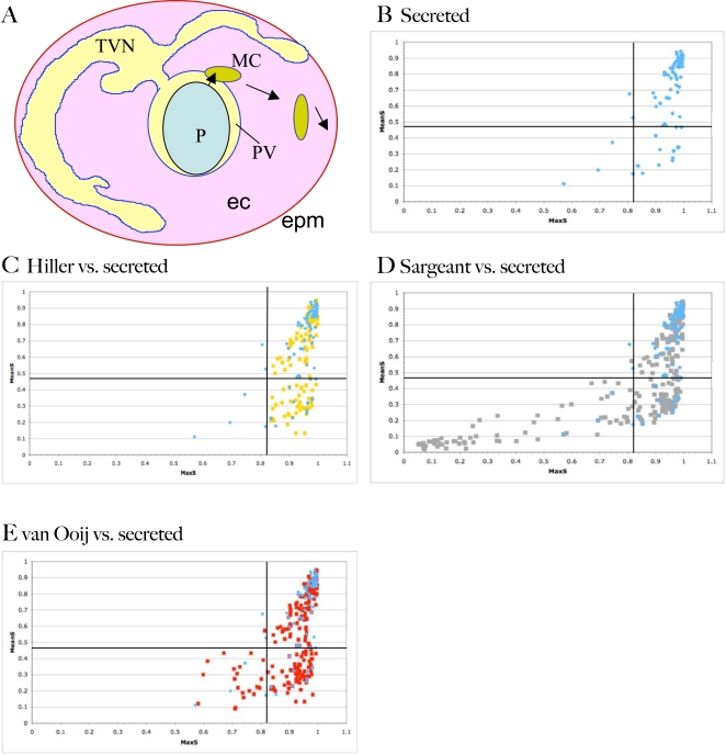
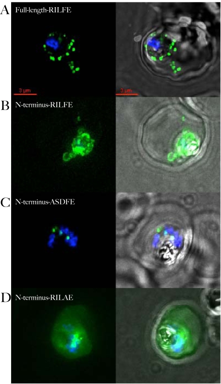
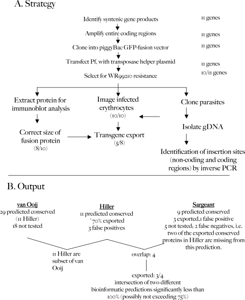
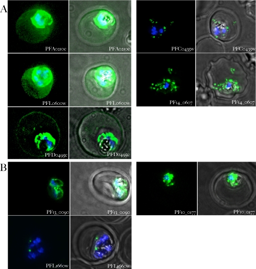
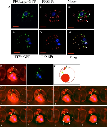
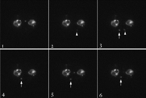
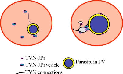
The malaria agent Plasmodium falciparum is predicted to export a "secretome" of several hundred proteins to remodel the host erythrocyte. Prediction of protein export is based on the presence of an ER-type signal sequence and a downstream Host-Targeting (HT) motif (which is similar to, but distinct from, the closely related Plasmodium Export Element [PEXEL]). Previous attempts to determine the entire secretome, using either the HT-motif or the PEXEL, have yielded large sets of proteins, which have not been comprehensively tested. We present here an expanded secretome that is optimized for both P. falciparum signal sequences and the HT-motif. From the most conservative of these three secretome predictions, we identify 11 proteins that are preserved across human- and rodent-infecting Plasmodium species. The conservation of these proteins likely indicates that they perform important functions in the interaction with and remodeling of the host erythrocyte important for all Plasmodium parasites. Using the piggyBac transposition system, we validate their export and find a positive prediction rate of approximately 70%. Even for proteins identified by all secretomes, the positive prediction rate is not likely to exceed approximately 75%. Attempted deletions of the genes encoding the conserved exported proteins were not successful, but additional functional analyses revealed the first conserved secretome function. This gave new insight into mechanisms for the assembly of the parasite-induced tubovesicular network needed for import of nutrients into the infected erythrocyte. Thus, genomic screens combined with functional assays provide unexpected and fundamental insights into host remodeling by this major human pathogen.


Conflict of interest statement

The authors have declared that no competing interests exist.

Figures

Figure 1
Figure 1. Export of proteins from P. falciparum to the erythrocyte.
A) Schematic of an erythrocyte infected with Plasmodium falciparum. Shown are the parasite (P), the surrounding parasitophorous vacuole (PV), the tubovesicular network (TVN), Maurer's Clefts (MC), the erythrocyte cytosol (ec) and the erythrocyte plasma membrane (epm). Arrows indicate the two steps involved in export of parasite proteins to the erythrocyte, ER-based secretion to the PV followed by HT-dependent transport to clefts (short arrow) followed by cleft movement and subsequent protein delivery to the cytoplasm and membrane of the erythrocyte (long arrow). B) Distribution of MaxS and MeanS values of 82 P. falciparum proteins known to enter the secretory pathway. Proteins were identified from the literature and include proteins destined for the apicoplast, apical organelles, parasite plasma membrane, food vacuole, erythrocyte cytosol and erythrocyte plasma membrane (Table S1). MaxS and MeanS values of the N-terminal 100 amino acids were identified using the SignalP 2.0 server. Lines indicate the MaxS and MeanS cut-offs used by SignalP (0.82 and 0.47, respectively). Comparison of the MaxS and MeanS values of the secreted proteins in B) (shown in blue) with the Hiller secretome (yellow) (C), Sargeant secretome (grey) (D), and van Ooij secretome (red) (E). RIFINS and STEVORs are not represented in the plots.
Figure 2
Figure 2. Export of PF14_0607-GFP requires full-length protein.
A) Localization of full-length PF14_0607 fused to GFP. The fusion protein is clearly detected in the erythrocyte cytosol. B) Fusion of the wild type N-terminal 89 amino acids to GFP is retained within the PV. Presence of GFP in cytoplasmic loops in the absence of staining in the erythrocyte cytoplasm indicates that the protein is secreted to the PV but not exported into the erythrocyte. C) Replacement of the HT-motif with unrelated sequence leads to retention of the protein inside the parasite. The punctate staining of the mutant fusion protein is reminiscent of apicoplast localization. D) Replacement of phenylalanine at position 4 in the HT-motif with an alanine residue allows the fusion protein to be exported. For each construct, the sequence of the HT-motif, or the sequence replacing it, is shown in the upper left hand corner of the panel. The overlap of GFP and Hoechst staining is shown in the left-hand panels, overlap with the phase contrast image is shown in the right-hand panels. All images are composites of multiple consecutive optical sections.
Figure 3
Figure 3. Outline for the validation of secretome predictions.
A) Strategy for expression and analysis of syntenic gene products. Numbers on the right-hand side and in parentheses indicate the number of genes that were positive over the number of genes tested. B) Output of data obtained from part A and corresponding analyses in context of Hiller, Sargeant and van Ooij secretomes.
Figure 4
Figure 4. Validation of export of syntenic gene products.
A) Syntenic gene products exported to the erythrocyte. Parasites expressing the gene indicated in each panel fused to gfp were stained with Hoechst 33342 and examined 18–36 hours after infection. Panels on the left-hand side show the overlap of the Hoechst 33342 staining and the GFP fluorescence, the panels on the right show the Hoechst 33342 staining, the GFP fluorescence and a phase contrast image of the infected cell. B) Syntenic gene products not exported to the erythrocyte. Samples were treated as in A. Notice the different patterns of distribution of the different gene products. All images are composites of multiple consecutive optical sections.
Figure 5
Figure 5. PFC0435w is a junctional protein of the TVN.
A) Relative distribution of PFC0435w-GFP (i) or HTTM-GFP (ii) and Maurer's clefts marked by PfSBP1 (ii, iv) in P. falciparum infected erythrocytes.The cells were also stained with Hoechst 33342 (blue) to visualize the DNA of the parasite. Panels i, ii and iv, v are single optical sections, whose merge is shown in iii and vi respectively. Scale bar, 2 µm. B–D) Association of PFC0435w-GFP with membrane buds and TVN structures B. Zero degree projection of an erythrocyte infected with a PFC0435w-GFP-expressing parasite stained with Rhodamine B. The numbers indicate the large circular TVN structure (1), and PFC0435w-GFP associated with other parts of the TVN (2 and 3). C) Cartoon representation of the infected cell in panel B. D) Optical sectioning of the infected erythrocyte shown in panel B. Distance between sections is 0.2 µm. Note the progression of PFC0435w-GFP staining from the PVM (sections 1–4) to the circular Rhodamine B-stained structure in the erythrocyte (sections 4–8). Arrows indicate PVM buds colocalizing with PFC0435w-GFP. Arrowhead indicates TVN connections between two large loops.
Figure 6
Figure 6. Still images from Video S1.
Images shown are the first six of the video (31 total), taken 1 second apart, with an exposure time of 180 msec. Note the appearance and disappearance of a vesicle from the plane of focus, as indicated with the arrow head, and the slower movement of a vesicle within the plane of focus, indicated by the arrow.
Figure 7
Figure 7. Model for action of conserved exported protein TVN-JP1 in the formation of the TVN.
In early (ring) stages (left-hand panel), TVN-JP1 is exported and present in small vesicles, which may emerge from the PVM and remain tethered to the PVM. At later stages (right-hand panel), TVN-JP1 is present in a neck structure, connecting the loops in the TVN to the PVM, as well as at sites interconnecting parts of the TVN, and on buds of the PVM.

Similar articles

Cited by

References

    1. Suwanarusk R, Cooke BM, Dondorp AM, Silamut K, Sattabongkot J, et al. The deformability of red blood cells parasitized by Plasmodium falciparum and P. vivax. J Infect Dis. 2004;189:190–194. - PubMed
    1. Miller LH, Usami S, Chien S. Alteration in the rheologic properties of Plasmodium knowlesi-infected red cells. A possible mechanism for capillary obstruction. J Clin Invest. 1971;50:1451–1455. - PMC - PubMed
    1. Cooke BM, Mohandas N, Coppel RL. The malaria-infected red blood cell: structural and functional changes. Adv Parasit. 2001;50:1–86. - PMC - PubMed
    1. Desai SA, Bezrukov SM, Zimmerberg J. A voltage-dependent channel involved in nutrient uptake by red blood cells infected with the malaria parasite. Nature. 2000;406:1001–1005. - PubMed
    1. Staines HM, Alkhalil A, Allen RJ, De Jonge HR, Derbyshire E, et al. Electrophysiological studies of malaria parasite-infected erythrocytes: current status. IntJ Parasitol. 2007;37:475–482. - PMC - PubMed

Publication types

LinkOut - more resources